Seminar Series Archive

2023 | 2022 | 2021 | 2020 | 2019 | 2018 | 2017 | 2016

The MU-MCW BME Graduate Seminar Series invites local, regional and national speakers to present current or novel biomedical research and recent innovation to graduate students in the MU-MCW Department of Biomedical Engineering. Below, view a complete list of this program's past speakers.


For a list of upcoming speakers, view the Graduate Seminar Series page. For further information on current or past speakers, please contact Dr. Tanya Onushko.


Past Speakers


Fall 2023

Don Anderson, PhD  September 15, 2023

Don Anderson is the Richard and Jan Johnson Chair in Orthopedic Biomechanics, Professor, and Vice-Chair of Research in the Department of Orthopedics & Rehabilitation at the University of Iowa. He holds a BSE in Biomedical Engineering, as well as an MS and PhD in Mechanical Engineering, all earned at the University of Iowa. Dr. Anderson has 30+ years of experience with image analysis, computer modeling, and computational stress analysis in musculoskeletal applications. Dr. Anderson's primary research focus throughout his career has been on articular joint biomechanics, specifically studying the mechanical relationship between joint injury and the subsequent development of post-traumatic osteoarthritis. All of his post-graduate career has been spent working in a clinical orthopedic setting, which has guided his work toward informing and influencing patient care.

Learn more about Dr. Anderson


Post-Traumatic Osteoarthritis Risk from Pathomechanics: Supporting Studies and New Intervention Strategies

The long-term goal of our research is to forestall post-traumatic osteoarthritis (PTOA), the disabling condition that often develops after joint injuries like an intra-articular fracture (IAF) of the tibial plafond. PTOA is one of the leading causes of mobility-related disability, affecting approximately 5.6 million individuals in the U.S. alone. PTOA leads to permanent disability in nearly 30% of individuals having sustained an IAF, with those of the foot and ankle being the most disabling. The impairment associated with ankle OA is comparable to that caused by end-stage kidney disease or congestive heart failure. The vast majority of ankle OA is post-traumatic, with tibial plafond IAFs often leading to disabling PTOA within two to five years. As a result, patients with ankle injuries provide an ideal population in which to study this degenerative pathway so that we can optimize treatment. We have developed patient-specific precision medicine approaches to predict PTOA risk in the ankle using CT-based measures of pathomechanical factors associated with IAFs (fracture severity and elevated contact stress post-treatment) of the tibial plafond. A primary objective of the group’s present work is to enable the use of these innovative methods for assessing IAFs to better inform patient care and to guide future clinical trials of new therapies directed at mitigating or arresting the environment that triggers progressive joint degeneration.


Christian Kastrup, PhD  September 29, 2023

Dr. Christian Kastrup is a Professor in the Department of Surgery at MCW, a Senior Investigator with Versiti Blood Research Institute, and a Secondary Faculty member in the MU-MCW Joint Department of Biomedical Engineering.

Learn more about Dr. Kastrup


RNA and Lipid Nanomedicines to Control Bleeding and Thrombosis

Bleeding and thrombotic disorders are common, affecting hundreds of thousands of North Americans, often due to an imbalance between the formation (coagulation) and destruction (fibrinolysis) of blood clots. Modulating fibrinolysis instead of coagulation is an alternate clinical approach for patients with bleeding and thrombotic disorders. However, only a small number of medications can influence pro- and anti-fibrinolytic proteins and enzymes, and they are short-acting. We developed a library of RNA and lipid nanoparticle (LNP) agents targeting the synthesis of pro- and anti-fibrinolytic proteins in vivo to modulate fibrinolysis long-term. Encapsulating mRNA and siRNA agents in LNP enabled delivery to the liver and bone marrow, where many pro- and anti-fibrinolytic proteins are synthesized. siRNA mediates gene silencing by degrading the target mRNA, resulting in long-term depletion of the corresponding protein in blood plasma for weeks to months. These mRNA-LNP can also be delivered to platelets, which allows further control of blood proteins. Administering pro- and anti-fibrinolytic RNA-LNP modulated fibrinolysis in vivo for weeks to months in small and large animal models of bleeding and thrombosis, the appropriate siRNA-LNPs corrected bleeding or thrombosis, showing promising therapeutic potential for humans.


Amy Lenz, PhD  October 13, 2023

Dr. Amy Lenz is a Research Assistant Professor in the Department of Orthopaedics at the University of Utah School of Medicine.

Learn more about Dr. Lenz

Learn more about Lenz Research Group


Foot and Ankle Biomechanics: Empowering Clinical Interventions with Advanced Technology to Study 3D Morphology and Kinematics

The foot and ankle is a complex structure of numerous articular relationships which operate to provide a stable base of support through active and passive tissue interactions.  Altered morphology can lead to injury, instability, pathological deformity, and osteoarthritis.  My lab’s goal is to characterize healthy, diseased, and post-surgical foot and ankle morphology and in-vivo function to improve clinical treatment of ankle pathologies leading to end-stage ankle osteoarthritis. Our recent studies have investigated the relationship between morphology and in-vivo function of the subtalar joint in patients that received a tibiotalar arthrodesis or total ankle replacement (TAR) surgery. Dynamic joint articulation measurements, such as joint space distance, coverage, and congruence can be investigated in combination with morphology analyses using statistical shape modeling  and biplane fluoroscopy kinematics to investigate the form and function relationship occurring at the subtalar joint following surgical intervention. Our ongoing studies highlight the complexity of the foot and ankle, the value of a robust 3D analyses, the utility of in-vitro robotic experiments, and the necessity to further investigate interactions of function and morphology to clinically evaluate flatfoot deformity, osteoarthritis, and injury mechanisms.


Joseph T. Barbieri, PhD  October 27, 2023

Dr. Joseph T. Barbieri is a Professor in the Department of Microbiology and Immunology and is the Associate Director of the Medical Scientist Training Program at MCW. 

Learn more about Dr. Barbieri


Bo Wang, PhD  November 3, 2023

Dr. Bo Wang is an Assistant Professor in the MU-MCW Joint Department of Biomedical Engineering and the Director of the Tissue Regenerative Engineering (TRE) Lab.

Learn more about Dr. Wang


Application of Human Amniotic Membrane in Tissue Engineering

Human amniotic membrane (HAM) is the innermost layer of  the placenta, which is a thin and semi-transparent membrane and contains various collagenous, non-collagenous proteins, and growth factors. HAM has many unique biological properties, including anti-inflammation, low immunogenicity, anti-fibrosis, and promotion of epithelialization, that make it an ideal biomaterial for such clinical applications as ophthalmic, abdominal, dental, plastic surgeries, and wound healing. 

In our laboratory, we've successfully developed a method for decellularizing HAM, effectively removing cellular components while preserving crucial extracellular matrix (ECM) constituents, resulting a decellularized amniotic membrane (DAM). We have investigated the use of DAM as a wound dressing material for repairing several types of tissue injuries, including wounds in oral cavity, bone, liver, and muscle. Furthermore, we've explored the use of DAM in the fabrication of small vascular grafts, and these DAM-based grafts have demonstrated exceptional material stability, biocompatibility, and long-term patency when employed in vascular transplantation surgery. 

Monica Rosenberg, PhD  November 17, 2023

Dr. Monica Rosenberg is an Assistant Professor in the Department of Psychology at the University of Chicago. 

Learn more about Dr. Rosenberg

Characterizing attention dynamics with functional brain dynamics

Although maintaining attention to a task at hand is crucial to navigating daily life, our attentional state waxes and wanes. Despite our best efforts, fluctuations in focus are ubiquitous during psychological tasks and everyday activities, such as watching movies and listening to lectures. What brain systems predict these attentional state changes? Do similar neural dynamics underlie attentional dynamics regardless of what we attend to, or do associations between neural and attentional dynamics vary by context? To address these questions, I will first provide evidence that a common functional brain network predicts changes in attention task performance on time scales from minutes to months in independent datasets. I will then show evidence from a deep imaging study measuring attention fluctuations in controlled and naturalistic tasks. Latent state analysis revealed that a common brain state consistently predicted periods of inattention, but different brain states occurred when participants were attentive to the tasks and movies. Thus, associations between brain-state dynamics and attention dynamics are both context-general and context-specific: whereas a brain state associated with suboptimal attention is shared across contexts, the brain state optimal for focus varies with cognitive and attentional demands. 


Ken Tichauer, PhD  December 1, 2023

Dr. Kenneth Tichauer is an Associate Professor and the Associate Chair of Graduate Affairs in the Department of Biomedical Engineering at Illinois Institute of Technology.

Learn more about Dr. Tichauer


Quantitative methods in fluorescence guided surgery and retinal imaging

The focus of this presentation will be on highlighting methods for extracting quantitative physiological and biomolecular parameters from contrast enhanced imaging approaches. Parameters of interest will include blood flow, vascular permeability, cell-surface receptor concentrations, and other drug targets. Several preclinical and clinical applications will be covered, including surgical margin assessment in head and neck cancer tumor resection, rapid and noninvasive lymph node biopsy, early prediction of diabetic retinopathy, and in vivo monitoring of drug binding.


Spring 2023   

Expand all   |   Collapse all  

Colin Burnett, MD  January 20, 2023

Dr. Colin Burnett is a graduate of the University of Iowa Roy J. and Lucille A. Carver College of Medicine and a current Electrophysiology Fellow at the Medical College of Wisconsin. 

Learn more about Dr. Burnett


Putting the Heat Back into Calorimetry

Obesity continues to worsen along with its comorbid conditions, and the limited efficacy of current pharmacotherapies necessitates reevaluation of the most common method of metabolic rate measurement: gas respirometry. Direct calorimetry overcomes the limitations of gas respirometry to reveal unappreciated changes in metabolism. Designing better calorimeters will become necessary to advance the understanding of the pathophysiology of obesity and to create more effective treatments of obesity.


Austin Stellpflug, PhD Candidate  February 3, 2023

Austin is a doctoral candidate and research assistant in Dr. Bo Wang's Tissue Regenerative Engineering Laboratory (TRE Lab).

Learn more about the TRE Lab


Development of a New Generation of Nanoparticles with Acellular Porcine Bone for Regenerative Engineering and Orthopedic Therapy

The increasing prevalence of bone-related diseases and physiological conditions has posed a significant health challenge, particularly to an aging society and those with bone-disease-related conditions. As an important tool of nanomedicine, Nanoparticles (NPs) have been extensively studied in areas including disease diagnosis and therapy, biomedical imaging, and drug and gene delivery, in large part due to their low toxicity, controllable physical stability, and tailorable characteristics for surface binding and encapsulation of drugs or molecules of interest. Although there are currently no FDA approved NP options for clinical orthopedic treatment, scientists are developing new NPs for improving the safety, diagnostic, and therapeutic efficiency that will address potential risks associated with NP use including elevated risks of low biocompatibility and drug delivery efficiency, unwanted accumulation in non-targeted organs, and a high chance for toxic side effects. Our lab has developed a novel type of multifunctional bone-based nanoparticle (BPs) using the decellularized extracellular matrix (dECM) of porcine bone tissue designed for orthopedic use. In this talk, I will discuss our current research using the BPs including 1) Local application of BPs for In vivo bone regeneration and healing, 2) Encapsulation of Indocyanine Green (ICG) for use with Near-Infrared Spectroscopy Imaging (NIR) in both local and systemic applications, and 3) Modification for bone-targeting systemic delivery with in situ, real time monitoring capability.


Hernan Rey, PhD  February 17, 2023

Dr. Rey is an assistant professor in the Department of Neurosurgery at the Medical College of Wisconsin and the Marquette-MCW Joint Department of Biomedical Engineering. He graduated in Electronic Engineering from the University of Buenos Aires, where he also obtained his PhD. He joined the University of Leicester (UK) in 2010 to perform his postdoctoral studies, and in 2012 he was awarded a Special Training Fellowship in Biomedical Informatics from the Medical Research Council (UK).

Learn more about Dr. Rey


Recording Neural Activity 24/7 at Different Scales in the Human Brain: A Unique Opportunity to Study Mechanisms Underlying Memory and Epilepsy

Epilepsy affects about 1% of the population worldwide, causing mortality, morbidity and reducing quality of life. When epilepsy becomes refractory (i.e., pharmacologically resistant), it is common to implant (macro) electrodes to record intracranial electroencephalogram (iEEG) in candidates identified for a surgical solution. In this talk, I will show you how we can apply modern technologies to simultaneously record from the human brain at different scales: from iEEG with clinical electrodes, to field potentials and single neuron activity from microwires protruding from the clinical probe. In fact, we are now able to record 24/7 for about a week, while the patient undergoes long-term monitoring to identify the epileptogenic zone (EZ), the area of the brain that is necessary for the appearance of seizures. During this time, the patient can engage in behavioral tasks while the neural signals are being recorded. In my lab, we are developing a research program that would allow to use this setup to answer important questions in clinical and cognitive neuroscience. The cognitive studies will allow us to unravel the building blocks of episodic memory and the associated neural mechanism, but will also provide us a platform to study other brain processes like perception, attention, language, and decision making. On the clinical research, we will search for new biomarkers to improve diagnosis and treatment in epilepsy. To achieve this, we rely on developing and applying methods to improve acquisition and analysis of neurophysiological data, based on concepts from signal processing and machine learning.


Marcella Vaicik, PhD  March 24, 2023

Dr. Vaicik is an assistant professor of Biomedical Engineering within the Armour College of Engineering at the Illinois Institute of Technology. She earned her BS in Chemical Engineering from Purdue, and earned her MS in Bioengineering from the University of Illinois at Chicago. She completed her PhD at the Illinois Institute of Technology and her postdoctoral research at the Department of Veterans Affairs, Edward Hines Jr. VA Hospital, in Illinois.

Learn more about Dr. Vaicik


Novel Therapeutic Targets and Development of Drug Delivery Systems for Use in Adipose Tissues Abstract

Adipose tissue (fat), the largest endocrine organ in the body, is comprised of adipocytes as the main cell type surrounded by specialized extracellular matrix (ECM). The ECM is a network of proteins that regulate adhesion, migration, proliferation, apoptosis, or differentiation in adipose tissue. Recent research from our group and others has emerged with new therapeutic targets in the ECM to instruct cell behavior in adipose tissue. Additional drug delivery systems are needed to realize the potential of these novel therapeutic targets. A major challenge in adipose tissue is that Adipocyte cells are difficult to transfect cells making drug delivery challenging. Novel polymeric hydrogel nanoparticles in inverse mini-emulsion are in development to achieve long-term drug delivery into hard to transfect cells. Engineering strategies to successfully identify and target adipocytes and ECM in adipose tissue has the potential to deliver drugs to treat metabolic disorders, type II diabetes and obesity.


Song Hu, PhD April 14, 2023

Song Hu’s research focuses on the development of cutting-edge optical and photoacoustic technologies for high-resolution structural, functional, metabolic and molecular imaging in vivo and their applications in neurovascular disorders, cardiovascular diseases, regenerative medicine, and cancer. 

Learn more about Dr. Hu


Light + Sound: Peering into Brain Function and Metabolism

Exploiting the optical absorption contrast of blood hemoglobin, photoacoustic microscopy (PAM) is an emerging technology for label-free imaging of the microvasculature, which plays an essential role in supplying oxygen to the biological tissue and maintain the metabolic activity in vivo. The multi-parametric PAM developed in Dr. Hu's lab enables, for the first time, comprehensive and quantitative characterization of the microvascular structure, function, and associated tissue oxygen metabolism at the microscopic level. In this seminar, Dr. Hu will present their latest progress on the development of PAM and the integration of PAM with other intravital light microscopy techniques for studying brain function and energy metabolism.


Yubing Tong, PhD  April 21, 2023

Dr. Yubing Tong is a Senior Research Investigator at the University of Pennsylvania Center for Undergraduate Research and Fellowships.

Learn more about Dr. Tong 


MR Image Segmentation & Analytics to Characterize the Upper Airway Structure in Obese Children with Obstructive Sleep Apnea Syndrome (OSAS)

This aims to introduce Dr. Tong's work on Obstructive Sleep Apnea Syndrome (OSAS) within recent years. It will include upper airway segmentation and image analysis to characterize the upper airway's structure in patients with OSAS. The approaches of upper airway and surrounding object segmentation from static MRI and dynamic MRI will also be introduced. Previous approaches of non-Deep Learning (DL) using automatic anatomy recognition (AAR) for upper segmentation, and the recently developed hybrid intelligence-based segmentation, combining natural intelligence and artificial intelligence, will be introduced. The upper airway segmentation from dynamic MRI includes work using both non-DL and DL techniques. The image analysis part will briefly introduce our approach of extracting useful features/biomarker to distinguish patients with Obese OASAS versus those who are obese but non-OSAS. In general, this talk will include an anatomy introduction, technical perspectives of deep learning and non-deep learning approaches, as well as feature extraction and prediction.


Whitney Morelli, PhD  April 28, 2023

Dr. Morelli (Welch) completed her BS and MS degrees at the University of Tennessee, Knoxville, and earned her PhD in Exercise Physiology at the University of Wisconsin—Milwaukee. She completed her postdoctoral fellowship at Northwestern University Feinberg School of Medicine in the NCI-funded T32 training program in cancer prevention and survivorship. In 2019, she was appointed to a faculty position at Northwestern University Feinberg School of Medicine prior to joining the MCW faculty in 2022.

Learn more about Dr. Morelli


Development of Patient-Centered, Personalized Physical Activity Interventions Using Digital Health Technology

Dr. Morelli (Welch) is an exercise physiologist whose overall research goal is to increase physical activity in populations at high risk for being inactive, with an emphasis on preventing or managing chronic disease. Her research interests and expertise include creatively using digital health technology, such as wearable devices, to develop and test innovative physical activity interventions to manage or prevent chronic disease or physical function decline among high-risk populations (cancer survivors, older adults). Dr. Morelli is particularly interested in moving beyond the "one size fits all" physical activity prescription and developing tailored, personalized physical activity prescriptions for individuals based on their personal, social, emotional, and physical circumstances. This talk will explore the opportunities to collaboratively work at the intersection of behavioral medicine and biomedical engineering to improve the health and quality of life for those with chronic conditions. 


Fall 2022   

Expand all   |   Collapse all  

Alex Walsh, PhD  September 30, 2022

Dr. Alexandra Walsh is an Assistant Professor of Biomedical Engineering at Texas A&M University.  Her research interests include biophotonics, image analysis, and laser-tissue interactions. 

Learn more about Dr. Walsh


Label-free Quantitative Imaging of Cellular Metabolism via Fluorescence Microscopy 

Cellular metabolism, the process by which cells generate energy, is dysregulated in many diseases and pathologies including cancer, neurodegeneration, and diabetes. Current biochemical assays for metabolism are limited to either cell-destructive protocols, such as mRNA analysis, and/or measure readouts from collective cell populations, such as oxygen consumption assays.  Yet single-cell measurements of metabolism are important since cellular heterogeneity is known to drive disease progression, cancer metastasis, and resistance to therapies.  Fluorescence lifetime imaging of the metabolic coenzymes, reduced nicotinamide adenine (phosphate) dinucleotide (NAD(P)H) and oxidized flavin adenine dinucleotide (FAD), provides a label-free method to interrogate cellular metabolism. Both coenzymes, NAD(P)H and FAD, exist in either a free or protein-bound configuration, each of which has a distinct fluorescence lifetime.  Single-cell segmentation and analysis of fluorescence lifetime images allows metabolic measurements at the cellular level.  To facilitate cell-level analysis of fluorescence images, we are developing automated segmentation algorithms.  Additionally, we are creating and testing models for predicting metabolic phenotypes from fluorescence lifetime metrics.  Our applications of single-cell metabolic phenotyping include evaluating temporal responses of cancer cells to chemotherapy and characterizing macrophage phenotypes.


Justin Grobe, PhD  October 7, 2022

Dr. Justin Grobe moved to the Medical College of Wisconsin’s Department of Physiology in June 2019. He holds a secondary appointment in the Department of Biomedical Engineering, and serves as the founding director of the Comprehensive Rodent Metabolic Phenotyping Core facility. Before joining the faculty at MCW, Dr. Grobe was a tenured associate professor in the Department of Pharmacology at the University of Iowa.

Learn more about Dr. Grobe


The Brain Renin-Angiotensin System in Metabolic Control and Obesity & Introduction to the MCW Comprehensive Rodent Metabolic Phenotyping Core (CRMPC)

Roughly 71% of adult Americans are overweight or obese, and although people can lose weight with diet, exercise, drugs, and surgeries, the majority regain that weight within a few years. Increasing evidence indicates that this weight regain is due to adaptation (suppression) of resting metabolic rate (RMR). This seminar will describe both ongoing research in our laboratory to understand RMR adaptation, and related rodent metabolic phenotyping capabilities provided by the new MCW Comprehensive Rodent Metabolic Phenotyping Core (CRMPC).

Part 1:  Our research has identified a key role for the angiotensin II type 1 receptor (AT1), localized to a specific subtype of neuron within the hypothalamus that also expresses Agouti-related peptide (AgRP), in the normal, physiological, integrative control of RMR. Specifically, we have determined that in the lean state, AT1 in AgRP neurons couples through a Gi-mediated pathway to inhibit the cell and thereby ultimately disinhibit (stimulate) RMR. We have also discovered that obesity and RMR adaptation are associated with a disruption of this second-messenger signaling cascade, as AT1 receptors stop signaling through the Gi pathway and instead exhibit a “G protein switch” in which these receptors begin to signal via a Gq-mediated, stimulatory pathway. Finally, our work suggests that reactivation of the normal Gi signaling cascade within this neuron can restore RMR control even during obesity. We propose that this G protein switch within the AgRP neuron represents a molecular basis for the clinical manifestation of RMR adaptation during or following obesity.

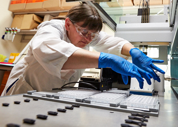
Part 2:  The new MCW CRMPC offers guided and fee-for-service access to advanced cardiometabolic phenotyping capabilities for rodents of various sizes (including mice and rats). Examples of major equipment offerings include a 16-chamber multiplexed home-cage phenotyping system (Promethion, Sable Systems International), a time-domain nuclear magnetic resonance body composition analyzer (LF110, Bruker), semi-micro bomb calorimeters (Parr), metabolic caging (Tecniplast), bioimpedance spectroscopy (ImpediVet), respirometry/indirect and direct calorimetry systems, an ashing oven, flame atomic absorption spectrophotometry, freezing point depression osmometry, and more (please see PMC8914175 for more details). The core additionally provides training, experimental design, best practice, data analysis, and statistical support.


Agrahara Bharatkumar, MD  October 14, 2022

Dr. Agrahara Bharatkumar is an associate professor of diagnostic radiology at the Froedtert and the Medical College of Wisconsin. 

Learn more about Dr. Bharatkumar


Artificial Intelligence in Radiology

Artificial intelligence (AI) is the computer science term used for methods that enable computers to perform tasks that normally require human intelligence. “Machine learning” is a subset of AI, and refers to the process by which computer algorithms “learn” from datasets in order to develop predictive models. The application of machine learning techniques to medical images has become a topic of great interest to researchers and practitioners alike. While traditional machine learning methods require hand-engineered feature extraction from inputs in order to point out the relevant features of the data on which predictive models are based, “deep learning” refers to a class of machine learning methods that learn what relevant features to extract directly from raw input data in order to use that data to develop predictive models. Deep learning models are multilayer artificial neural networks, loosely inspired by biologic neural systems, and they are gaining success and attracting interest in many domains, including computer vision, speech recognition, and natural language processing. With the advent of large datasets and increased computing power, deep learning methods can produce models with exceptional performance – that is, exceptional predictive capability – but without the need for feature engineering or data labeling. For computer vision tasks – or tasks by which computers derive information and understanding from images, convolutional neural networks (CNNs) have proven to be effective. Recently, several clinical applications of CNNs have been proposed and studied in radiology for classification, detection, and segmentation tasks. This seminar will review key fundamental concepts of AI, briefly describe emerging applications of AI in clinical radiology, and outline limitations and future directions in this field.


Peter LaViolette, PhD  October 28, 2022

Dr. LaViolette is an Associate Professor in the Department of Radiology at the Medical College of Wisconsin. His research focuses on creating and validating imaging techniques that improve patient outcome and treatment efficacy. 

Learn more about Dr. LaViolette


Radio-pathomic Mapping of Brain Cancer

The computational extraction of features from radiographic imaging of cancer is termed radiomics, and combining this data with tumor genetics is termed radio-genomics. Our research is taking this idea further by training predictive models with machine learning applied to pathology samples aligned to MRI. The results can be applied to imaging data from patients prior to intervention to produce quantitative ‘radio-pathomic’ maps of cancer characteristics. In this talk I will be discussing our ongoing efforts in brain cancer research where we are combining tissue from our brain bank with clinical imaging to detect tumor invasion beyond conventional margins defined by gadolinium contrast based methods. 


Kevin Dibbern, PhD  November 4, 2022

Dr. Kevin Dibbern is a postdoctoral fellow working with the labs at the Marquette University and Medical College of Wisconsin Orthopaedic & Rehabilitation Engineering Center (OREC). He has done previous work with foot and ankle weightbearing CT imaging, and is a recent doctoral graduate of the University of Iowa.

Learn more about the OREC


Automatable 3D Assessment of Weightbearing CT Data: Improving Diagnosis and Treatment in the Foot and Ankle

Until recently, standard diagnosis and treatment of foot and ankle pathologies were dependent on 2D X-ray measurements or non-weightbearing 3D imaging modalities like MRI and conventional CT. Over the past decade, weightbearing conebeam CT (WBCT) has become available to perform rapid weightbearing assessment of the foot and ankle. It affords low-radiation-dose 3D imaging of the lower extremity in functional positions. Combined with recent research efforts, it has taken over as the gold standard for identifying flat foot and other foot and ankle pathologies. However, new clinical measurements have still relied upon planar 2D interpretation of these complex 3D data.

Recently, machine learning has enabled automated 3D model creation from these scans. This has led to a unique opportunity to quantitatively examine the lower limbs in their functionally loaded positions without losing relevant features to planar interpretation. Such advancements are enabling early detection of subtle changes related to injury and deformity and optimizing patient specific care though improved understanding of the type and timing surgical intervention needed.


Brian Hoffman, PhD  November 18, 2022

Dr. Brian Hoffmann is the Study Director of Mass Spectrometry and Protein Chemistry at The Jackson Laboratory and former faculty of the Marquette-Medical College of Wisconsin Joint Department of Biomedical Engineering. 


Advances and Applications in Mass Spectrometry Imaging

In recent years there have been significant advances in high-resolution mass spectrometry imaging of tissues to measure the spatial distribution of lipids, small molecules, and peptides.  Using matrix-assisted laser desorption/ionization technology coupled with ion mobility, spectral images representing the distribution of analytes at high-resolution (5–10 um) across the entirety of a tissue section can be acquired.  These analyses also benefit from trapped ion mobility spectrometry (TIMS) to separate isobaric or isomeric analytes, allowing you to differentiate analytes when high mass resolution is not sufficient alone.  In this talk, I will be discussing advances in mass spectrometry imaging, sample preparation considerations, and provide examples of data acquired from multiple tissue systems (brain, kidney, lung and tumor) using our facilities’ Bruker timsTOF FLEX.  In addition, the post-processing data analysis required and the associated challenges will be discussed.  Lastly, the talk will explore data collected with our collaborator, Dr. Alison Kriegel (MCW), focused on the progression of changes in the lipid profile of mouse kidneys during polycystic disease.


Daniel Sem, JD, PhD  December 2, 2022

Dr. Daniel Sem is Vice Provost of Research and Innovation at Concordia University. He has terminal degrees in intellectual property law and biochemistry. He has special interest in pharmaceutical design and biochemistry.


Resources for Healthcare and HealthTech Startups in Southeast Wisconsin

Healthcare and HealthTech startups, coming largely from regional universities, have many needs. This talk will review resources connected to CTSI, as well as resources that assist startups in the areas of drafting business models, plans and investor pitches, corporate and legal assistant with forming a company and closing investor rounds, raising investor capital, types of investment instruments (e.g., SAFE, Convertible notes, value rounds), identifying talent for a startup, regulatory advice for drugs, devices and diagnostics, and networking to connect with other startups to identify synergies and partnerships. Resources discussed will include CTSI-AMPDNR (Accelerating Medical Products through Networked Resources), BioForward and HealthTech MKE, Catalyst Bioconsulting, NSF and NIH I-Corps, Bridge to Cures, WEDC, CTC, and the various angel investor groups, including Brightstar, MVP, Golden Angels and CU Ventures. Examples of startups that have navigated these steps will also be discussed.


Derek Kamper, PhD  December 9, 2022

Dr. Derek Kamper is an associate professor of Biomedical Engineering within the Joint Department of Biomedical Engineering at NC State and the University of North Carolina. His research focuses on  improvement of upper extremity function, especially for the hand, in cases of injury or disease. 

Learn more about Dr. Kamper


Addressing Functional Hand Limitations After Stroke

In the Hand Rehabilitation Lab, Dr. Kamper and his team seek to improve outcomes for individuals with neuromuscular impairments, especially those affecting control of the hand. The lab's strategy has been to increase understanding of sensorimotor control, to identify mechanisms of impairment, and to use this knowledge to develop new interventions. As an example, Dr. Kamper will present a completed clinical trial involving stroke survivors with chronic upper extremity deficits. After stroke, functional limitations often arise due to a seemingly paradoxical combination of involuntary motor unit hyperexcitability and diminished motor unit excitation during voluntary activation. The lab has attempted to address both phenomena through a multimodal intervention by administering cyproheptadine hydrochloride to reduce involuntary muscle activation and by instituting active training of muscle activation patterns with an electromyographically (EMG) driven hand exoskeleton and EMG-controlled “serious” computer games.


Spring 2022

Expand all   |   Collapse all  

Mercedes Rodriguez Celin, MD  January 28, 2022

Dr. Celin is an experienced pediatrician with a demonstrated history of working with children with motor disabilities, skeletal dysplasias and growth problems in pediatric hospitals. 

Learn more about Dr. Celin


Abstract

A Multicenter Study to Evaluate Pain Characteristics in Osteogenesis Imperfecta

The goals of this study were to investigate the characteristics of chronic pain in various osteogenesis imperfecta (OI) types, analyze association of pain with different clinical co-variates, and evaluate pain interference in activities. Cross-sectional analysis of data on adults and children enrolled in the Brittle Bone Disorders Consortium. Characteristics of chronic pain, demographic, clinical, and surgical co-variates were collected. Mobility outcomes were analyzed using Gillette Functional Assessment Questionnaire (GFAQ), Functional Mobility Scale (FMS), Brief Assessment Motor Function (BAMF), and 6-minute walk test (6MWT). Pain intensity, pain interference and limitation to participation were analyzed from PODCI and SF-12v2.


Alkis Hadjiosif, PhD  February 4, 2022

Alkis Hadjiosif is a Postdoctoral Fellow at Johns Hopkins University. He received his Diploma in Electrical and Computer Engineering from the National Technical University of Athens, Greece in 2008 and his PhD in Bioengineering from Harvard University in 2015 studying the influence of environmental variability in motor adaptation. His research aims to understand mechanisms of motor learning and motor control, in both healthy individuals and ones afflicted by motor disorders, and leverage this understanding to improve the assessment and rehabilitation of neurological impairment in the motor system.

Learn more about Dr. Hadjiosif


Abstract

Post-stroke Postural Abnormalities in the Arm and Their Relationship to Loss of Motor Control

Abnormal resting postures are one of the most common and widely recognizable motor symptoms after stroke. For example, the typical hemiparetic arm posture consists of flexion at the fingers, wrist, and elbow. This pattern appears to parallel the abnormal muscle synergies during active movement, such as the abnormal coupling of muscles that move the shoulder vs. the elbow joint. Whether these synergies are generated by the same mechanism as abnormal resting postures remains an open question; it might instead be that resting abnormalities are inactive during movement. Also unknown is the degree to which resting postural abnormalities influence active moving and holding.

Here we systematically assessed resting postural abnormalities in stroke patients. We found that patients exhibited abnormal postural forces at rest, which mirrored characteristics of abnormal synergies: for example, postural abnormalities were markedly lower when the arm was supported against gravity, and, critically, strongly related to the Fugl-Meyer scale, a measure based on abnormal synergies. These findings suggest a shared mechanism between resting abnormalities and abnormal synergies after stroke.

We then examined whether these resting postural abnormalities affected the motor control of active reaching in the same workspace. We did not find any systematic effects of resting postural forces either across patients with different resting posture magnitudes, nor between high- vs. low-resting postural force regions for the same patient. However, we found evidence suggesting that resting postural abnormalities might influence active holding control. We conclude that post-stroke deficits in posture and movement may be driven by separate mechanisms. Moreover, our findings identify arm support as a way to alleviate these resting postural abnormalities.


Jordan J. Williams, MD, PhD  March 4, 2022

Dr. Williams is an assistant professor within the Marquette-MCW Joint Department of Biomedical Engineering, and Director and Principal Investigator of the NEIMO Lab, which employs a combination of neurophysiology, optogenetics, viral gene therapy, and optical imaging techniques to develop novel neuroprosthetic and gene therapy approaches to alleviate motor deficits caused by conditions such as spinal cord injury or ALS. 

Learn more about Dr. Williams


Abstract

Peripheral Optogenetic Stimulation of Motor Activity for Spinal Cord Injury Rehabilitation

Electrical stimulation of muscle activity has been the mainstay approach to reanimating paralyzed muscle function in conditions such as spinal cord injury for decades.  However, this approach has traditionally been plagued by several drawbacks including an unnatural recruitment order of muscle fibers and early muscle fatigue that have limited its therapeutic ceiling for patients.  Conversely, peripheral optogenetic stimulation of muscle activity is a recently emerging approach that has shown promise in alleviating these drawbacks in addition to other unique advantages.  In this gene therapy technology, nerves are labelled with light-sensitive ion channels via virus injection that allow them to be stimulated with light.  However, modifying the biology of the peripheral nervous system in this manner (as opposed to only electrically interfacing with it) raises its own set of challenges including exposure to the immune system and long paths of retrograde virus travel after muscle injection in order to reach targeted motor neurons in the spinal cord.  In this talk, I will discuss some of my lab’s recent approaches in rodent and non-human primate models to characterize and overcome these hurdles to clinical translation.  In addition, I will describe our efforts to exploit previously underutilized advantages of peripheral optogenetic neuromodulation as well as to extend these viral transduction techniques to spinal cord injury models.


Roger Guillory, PhD  March 25, 2022

Roger Guillory II is an assistant professor of Biomedical Engineering at Michigan Tech with research interests including cellular and molecular interactions of biomaterials, metallic biocorrosion, biomaterial histopathology, biocompatibility, and degradable metals for cardiovascular implants.

Learn more about Dr. Guillory


Abstract

The interplay of Biodegradable Metal Performance and Physiological Microenvironments: Informing the Material Design of Advanced Cardiovascular Implants

Non-degradable metal stents are the current gold standard in treating patients with coronary artery disease. However, there is an interest in pursuing metal stents made from biodegradable metals such as magnesium or zinc, in an effort to minimize long term complications in patients. Metallic magnesium and zinc are prominent metal candidates for stenting applications, although both require advanced metallurgical and surface engineering in order to reach the appropriate biomechanical and biodegradation benchmarks. These engineering modifications while necessary, can cause shifts in the clinical performance of the materials. The link between biocompatibility, elemental additions, microstructural evolution, and biodegradation profiles remains elusive for biodegradable metals.  We have identified key metallurgical and biodegradation factors that influence the tissue response of advanced biodegradable metals in-vivo using a large-scale statistical histological approach. However, understanding the tissue response towards any implanted degrading vascular material is confounded by the complicated material and biological variables which contribute towards the final outcome. Presently, it is unclear how material biodegradation rate, implant derived metal cations, cellular injury, or cellular activity contribute to negative biologic reactions to the materials. To fill this knowledge gap, we have been investigating the bioactivity of biodegradable metal materials in-vitro in order to relate cellular events such as cell death, intracellular metal regulation, reactive species generation, with alloy composition and biodegradation characteristics. Our work demonstrates the need for a critical understanding of the biometal-biocorrosion-cellular/tissue response axis, in order to rationally design future degradable metal materials for cardiovascular applications. 


Gui Garcia, PhD  April 1, 2022

Dr. Garcia is an assistant professor of Biomedical Engineering within the Marquette University - Medical College of Wisconsin Joint Department of Biomedical Engineering. 

Learn more about Dr. Garcia


Abstract

Modeling Drug Delivery to the Nasal Passages with Nasal Sprays

Nasal sprays are widely used over-the-counter medications to treat sinusitis, allergic rhinitis, and nasal obstruction. The Food and Drug Administration (FDA) is interested in understanding how the intranasal distribution, absorption, and bioavailability of the aerosolized medication is influenced by factors such as interindividual variability in nasal anatomy, spray nozzle position in the nostril, and spray pump characteristics that vary among commercial products (e.g., initial droplet velocity, droplet size distribution, spray cone angle, etc.). In this presentation, I will summarize an ongoing project in the Airway Lab that is funded by the FDA to develop and validate computer models of intranasal distribution of nasal sprays. We will discuss how the computer models are validated with in vitro experiments in anatomically accurate, 3D printed nasal replicas.  To illustrate a practical application of the model, we will discuss how the nozzle position within the nostril affects the dose delivered to relevant target sites.


Janelle Cross, PhD  April 8, 2022

Janelle Cross received her doctorate degree in Biomedical Engineering from Marquette University with a Rehabilitation Biomechanics specialization. Her dissertation work focused on developing a biplanar fluoroscopic system for analyzing in vivo and ankle kinematics during gait.

Learn more about Dr. Cross


Abstract

Adolescent Baseball Pitching Biomechanics: Implications on Injury Risk and Performance

Joint forces and moments are important factors in determining the risk of injury to baseball pitchers. Due to the repetitive nature of pitching, adolescent baseball pitchers are at a high risk for sustaining injuries to the upper extremity (UE) joints. Two critical periods of high risk of injury have been identified in current literature: 1) shortly before the point where the arm reaches maximum external rotation, when shoulder internal rotation torque and elbow valgus torque are generated and 2) shortly after ball release, when shoulder compression force, posterior force and horizontal abduction torque are generated. A series of studies investigating adolescent baseball pitchers was conducted to assess relationships among injury history, hip clinical measures, spin rate and pitching biomechanics. Several associations were found that have indications for injury prevention and performance enhancement.


Josh Roth, PhD  April 22, 2022

Josh Roth is an Assistant Professor in the Departments of Orthopedics and Rehabilitation, and Mechanical Engineering at the University of Wisconsin-Madison. He earned a B.S. in Mechanical Engineering from California Polytechnic State University, and an M.S. and Ph.D. in Biomedical Engineering from the University of California, Davis. The mission of his research group, the Biomechanical Advances in Medicine (BAM) Lab, is to enhance personalized treatments of musculoskeletal injuries and disease. To fulfill this mission, he develops and applies translational technologies to measure joint and tissue biomechanics to enhance planning, execution, and evaluation in the clinical environment.

Learn more about Dr. Roth


Abstract

Development and Pre-Clinical Testing of Our Handheld Ligament Tensiometer           

Ligaments are important tissues that act like stiff springs connecting our bones at most joints. One key role of ligaments is to act as mechanical restraints to stabilize the joint and guide its motion. Injury, disease, and surgical intervention can disrupt a ligament’s tension-length relationship, which consequently disrupts its ability to stabilize the joint and guide its motion. Although clinicians attempt to account for these disruptions, there are currently no devices available to directly and non-invasively measure these changes in vivo. In this talk, I will describe the underlying theory and development progress of our ligament tensiometer, which is a hand-held sensor to directly and non-invasively measure ligament tension based on the shear wave propagation speed in the structure. I will also show two clinically relevant examples to demonstrate the promise of this sensor to enhance clinical decision making in total knee replacement.


Hyo Jung Jeong, PhD April 29, 2022

Dr. Jeong is a NIDILRR ARRT postdoctoral fellow at Marquette University. She received her Bachelor’s and Master’s degree in Physical Therapy at Yonsei University, South Korea. She has completed her Ph.D. training at Washington University in St. Louis, MO, in the Movement Science program, investigating foot and ankle biomechanics in people with diabetes. Her postdoctoral work investigates biomechanics of upper and lower extremities in children with hypermobile Ehlers-Danlos syndrome. She is particularly interested in understanding the rehabilitation of movement patterns over a lifespan.

Learn more about Dr. Jeong


Abstract

Biomechanical Phenotyping of Children with Hypermobile Ehlers-Danlos Syndrome  

Ehlers-Danlos syndrome (EDS) is a rare genetic connective tissue disorder that impacts 1/5000 people worldwide. Hypermobile EDS (hEDS) is the most common EDS subtype and the only subtype with an unknown genetic origin. Current diagnosis of hEDS often relies on clinical findings, which leads to delayed or misdiagnosis in hEDS and ultimately burdens the quality of life across the lifespan in children with hEDS. Therefore, characterizing a biomedical phenotype is necessary to understand the clinical features better and improve rehabilitation strategies for children with hEDS. The main goal of this talk is to share characteristics of movement phenotypes in children with and without hEDS.


Fall 2021   

 Expand all   |   Collapse all  

Regine Choe, PhD  September 03, 2021

Dr. Choe's research is focused on the development of diffuse optical methods for the clinical application of breast cancer diagnosis and therapy monitoring.

Learn more about Dr. Choe


Abstract

Non-invasive Hemodynamic Monitoring of Bone, Brain and Breast Using Diffuse Optics

Diffuse optical tomography (DOT) and diffuse correlation tomography (DCT) are non-invasive three-dimensional hemodynamic imaging techniques that can probe deep tissue using light sources in the near-infrared spectral window (650–950 nm). By modeling the photon propagation in tissue, one can quantify oxygenated hemoglobin, deoxygenated hemoglobin, water and lipid concentrations with DOT, and blood flow with DCT. These intrinsic physiological parameters have great potential to assess therapeutic efficacy of various treatments. In addition, the use of non-ionizing radiation and technologically simple, fast, inexpensive instrumentation makes diffuse optical and correlation tomography attractive for translational research.


Jim Hokanson, PhD  September 17, 2021

Dr. Hokanson's research interests include urologic function/dysfunction, electrical stimulation/neuromodulation therapies, signal processing and machine learning, clinical diagnostics, autonomic nervous system and organ physiology, and neural engineering.

Learn more about Dr. Hokanson


Abstract

Improving Incontinence Therapy Outcomes with Electrical Stimulation and Improved Diagnostics

Urinary incontinence is prevalent (estimated to impact over 40% of women over 40), has a high cost-burden, and negatively impacts quality of life. Most therapies for treating urgency urinary incontinence fail to make patients dry. One study demonstrated that upwards of 90% of patients fail to respond to drug therapy and another demonstrated that less than 20% of drug-refractory patients become completely dry after Botox or sacral neuromodulation therapies. In my talk I’ll highlight experimental work aimed at improving incontinence therapy using electrical stimulation, as well as statistical modeling aimed at improving diagnosis and therapy selection. With both sets of examples I’ll demonstrate how important science often happens along the journey rather than, or in addition to, getting to the destination. Implications for translating electrical stimulation studies from animals to people will also be discussed.


Mark Pierce, PhD  September 24, 2021

Dr. Pierce's lab develops novel optical imaging systems for applications ranging from basic biomedical research studies to clinical patient care. His lab also collaborates extensively with clinical partners in surgery, radiation oncology, pathology, and image analysis.

Learn more about Dr. Pierce


Abstract

Short-wave Infrared Optical Imaging in Small Animal Models of Cancer 

Short-wave infrared (SWIR) light has several advantages for biomedical optical imaging applications.  SWIR light (900–1700 nm wavelengths) scatters less within tissue than near-infrared or visible light, resulting in sharper images.  Tissue autofluorescence is negligible in the SWIR region, minimizing background light and improving contrast.  Our lab is developing rare-earth doped nanocomposites as imaging contrast agents which generate light at SWIR wavelengths.  Encapsulation within an albumin shell renders these nanocomposites biocompatible, while also providing binding sites for molecular targeting ligands.  We have used these nanocomposites to label tumors in small animal models of breast and ovarian cancer, toward potential applications in pre-clinical drug screening, tracking of micrometastatic lesions, and image guided surgery.  This presentation will discuss challenges and opportunities in imaging these SWIR-emitting nanocomposites across macro and microscopic spatial scales. 


Bin Jiang, PhD  October 01, 2021

Dr. Jiang is an assistant professor of biomedical engineering and surgery. Their research interests include vascular regeneration, stem cells, biomaterials, and tissue engineering.

Learn more about Dr. Jiang


Abstract

Autologous Vascular Regeneration for Peripheral Artery Disease with Induced Pluripotent Stem Cells

The clinical translation of stem cell-based therapies for the treatment of peripheral artery disease (PAD) is currently hindered by the lack of a defined cell source, sub-optimal delivery strategies, and unknown fate of administered cells. The advent of induced pluripotent stem cells (iPSCs) offers an exceptional opportunity for regenerating functional tissues for various diseases, including PAD. Nuclear imaging techniques together with reporter gene transgenic expression provide a highly sensitive, non-invasive tool to monitor the fate of viable transplanted cells in vivo. Furthermore, advanced functional biomaterial scaffolds that can deliver stem cells to the targeted tissues/organs and promote stem cell survival, differentiation and integration to host tissues may potentially transform the clinical outcome of stem-cell based regenerative therapies. In this seminar, I will cover our recent efforts in (1) generating autologous iPSC derived vascular endothelial cells (iPSC-ECs) as an autologous cell source for PAD patient vascular regeneration, (2) demonstrating non-invasive cell tracking in vivo using a clinical applicable non-invasive imaging technique, and (3) promoting long-term cell survival with advanced biomaterial design.  The hypotheses are: (1) the survival and biodistribution of iPSC-ECs expressing human sodium iodide symporter (hNIS) can be tracked non-invasively in vivo via Single Photon Emission Computed Tomography /Computed Tomography (SPECT/CT) without affecting EC functions; and (2) an antioxidant cell engraftment niche that promotes cell adhesion and spreading of iPSC-ECs, and supports vascular regeneration will reduce oxidative cell damage, improve cell engraftment and survival rate, and support limb revascularization. This study is essential to help establish optimized cell manufacturing protocols, develop cell preservation agents for limb revascularization, and develop cell tracking procedures to offer additional treatment options for PAD patients.


Alister Bates, PhD  October 08, 2021

Dr. Bates' research focuses on human airways and how they change with various disease conditions, specifically airway behavior in children with obstructive sleep apnea (OSA) and premature babies born with tracheomalacia (TM) and congenital abnormalities. 

Learn more about Dr. Bates


Abstract

Understanding Airway Movement Through CFD Simulations of Respiratory Airflow Based on Cine MRI 

Several respiratory diseases are caused by collapse and obstruction of the airway. In obstructive sleep apnea (OSA), this collapse occurs behind the tongue or soft palate, or in the larynx. In tracheomalacia, the trachea collapses. This talk will focus on both conditions; OSA in pediatric patients and tracheomalacia in neonates who are born prematurely. CFD simulations of respiratory airflow calculate many metrics that are clinically useful in understanding these conditions such as localized resistance, work of breathing, and pressure differences. However, to calculate these parameters in a moving airway, the airway motion must be realistically included in the simulation’s boundary conditions. One way to do this is to image airway motion using cine MRI, which provides a 3D airway image several times per second. The motion between each image can then be calculated via image registration and applied as a moving boundary condition. 

In OSA, CFD simulations with prescribed wall motion can be used to assess airway muscle tone and function by comparing how the airway wall moves with the aerodynamic pressures acting on the wall. Where the wall moves in a manner that cannot be explained by the pressure acting on it, muscular activity is the most likely cause of that motion. Given that the upper airway is made of highly dynamic structures such as the tongue, soft palate, and epiglottis, mapping muscular activity is an important consideration in understanding airway collapse in OSA. 

In neonates who are born prematurely, patients suffer abnormalities of both the lung parenchyma and the airway, in the form of tracheomalacia. Moving wall respiratory CFD simulations, allow the effect of the tracheomalacia to be calculated separately from the lung parenchymal issues which leads to individualized treatment plans to be designed for each baby. 


Daniel Beard, PhD  October 15, 2021

Dr. Beard is a Professor in the Department of Molecular and Integrative Physiology and holds affiliate appointments in Biomedical Engineering and Emergency Medicine. His laboratory is focused on systems engineering approaches to understanding the biophysical and biochemical operation of physiological systems. 

Learn more about Dr. Beard


Abstract

Metabolic and Mechanical Determinants of Reserve  Cardiac Power Output (What Determines Exercise Capacity?)

Changes in the myocardial energetics associated with aging and heart failure—reductions in creatine phosphate/ATP ratio, total creatine, and ATP—mirror changes observed in failing hearts compared to healthy controls. Similarly, both aging and heart failure are associated with significant reductions in cardiac performance and maximal left ventricular cardiac power output compared with young healthy individuals. Based on these observations, we hypothesize that reductions in the concentrations of cytoplasmic adenine nucleotide, creatine, and phosphate pools that occur with aging and age-associated disease impair the myocardial capacity to synthesize ATP at physiological free energy levels and that the resulting changes to myocardial energetic status impair the mechanical pumping ability of the heart. To test these hypotheses, to determine the potential impact of reductions in key myocardial metabolite pools in causing metabolic/energetic and cardiac mechanical dysfunction, we analyzed data on myocardial mechanics and energetics during voluntary exercise using a multiscale model of cardiac metabolism and mechanics.  Model simulations support the hypotheses and  provide a novel theoretical/computational framework for further probing complex relationships between the energetics and performance of the heart with aging.


David Cormode, PhD  October 29, 2021

Dr. Cormode's research focuses on the development of novel and multifunctional nanoparticle contrast agents for medical imaging applications. A current major focus is the development of gold and bismuth nanoparticles as contrast agents for computed tomography (CT). 

Learn more about Dr. Cormode


Abstract

Inorganic Nanoparticles for Multi-energy X-ray Imaging and Therapeutics  

X-ray based imaging has been used in medicine for the past 120 years. Despite the age of this technology, we are in an era of rapid innovation in x-ray imaging methods. In particular, multi-energy x-ray imaging (such as dual energy mammography) is becoming widely available to patients, and is providing benefits such as more accurate cancer detection. These new imaging technologies require novel contrast agents that are specifically aligned to them, to allow high contrast generation. The development of such novel contrast agents will be described. For example, silver-based nanoparticles that provide improved contrast in dual energy mammography and are highly biocompatible will be discussed. Specific and multiplexed detection of contrast agents such as gold nanoparticles with spectral photon-counting computed tomography will be presented. These agents are designed for clinical translation and so their biodegradation and excretion will be discussed. Applications of these agents/systems for tumor detection, vascular imaging and cell tracking will be described. Moreover, the use of these nanoparticles as anti-cancer and anti-biofilm agents will be reported. 


Christopher Pawela, PhD  November 05, 2021

Dr. Pawela was awarded a Ph.D. in Biophysics from the Medical College of Wisconsin in 2008. He joined the MCW faculty immediately upon graduation with research interests that include brain connectivity and neural plasticity.

Learn more about Dr. Pawela


Abstract

Neuroaugmentation in the Context of Peripheral Nerve Injury and Repair

Extremity injuries involving peripheral nerve damage are a significant medical risk for the modern warfighter. Nerve damage can result from blunt, penetrating, or compression injuries caused by explosions, projectiles, vehicular accidents, or other duty related events. Current treatment of peripheral nerve injuries by direct end-to-end microsurgical repair and nerve grafts/conduits is typically followed by incomplete recovery of sensorimotor function, abnormal phantom sensations, neuropathic pain, and muscle atrophy that often lead to permanent disability in veterans. Minimal advancement of treatment has been achieved in the last 30 years. To address this need, our novel treatment approach provides continuous electrical stimulation to the injured peripheral nerve immediately after nerve repair surgery through a bipolar nerve cuff electrode placed proximal to the repair site. This is based on our central hypothesis that the loss of normal sensory input to the central nervous system (CNS) leads to disordered neuroplasticity, and that surrogate activity provided by electrical stimulation of the injured nerve proximal to the injury can prevent the loss of connectivity between the CNS and the peripheral nervous system that otherwise leads to dysfunction. Our methodology is technologically similar to spinal cord stimulators (1968) and cardiac pacemakers (1958) which have been used clinically for over 50 years. In this seminar, data will be presented from pilot studies performed in an animal model that replicates many key features of clinical injury and rehabilitation. We have successfully applied our treatment approach in a rodent model following complete median nerve transection and repair. Our pilot studies, which were funded by a completed VA SPiRE grant, demonstrate a significant improvement in functional magnetic resonance (fMRI) measured brain response to median nerve stimulation, as well as skilled limb reaching performance as quantified by an automated behavioral device. Early pilot data also points to improvement in peripheral nerve regeneration after our treatment is applied, as measured by nerve conduction studies. Finally, we will discuss the optimization of treatment effectiveness of our novel methodology and future studies introducing impediments to regeneration that reflect clinical circumstances, such as interposed nerve grafts or conduits. The impact of various electrical stimulation patterns in neuroaugmentation will be discussed. Our goal is to first develop foundational work in animal models and proceed to eventual clinical translation for enhancing rehabilitation of injured veterans.


Harkamaljot "Rocky" S. Kandail, PhD  November 12, 2021

Dr. Kandail is interested in Computer-Aided (Biomedical) Engineering with particular emphasis on the amalgamation of clinical imaging techniques such as computed tomography and magnetic resonance imaging with computational cardiovascular biomechanics. He works to elucidate the role structural deformations and haemodynamics play in cardiovascular disease development and progression, along with designing novel biomedical devices to treat such diseases. Current approaches include computational fluid and solid mechanics along with fluid-structure interactions.

Learn more about Dr. Kandail


Abstract

Fluid-Structure Interaction Simulations for Paediatric Heart Valves: Challenges, Solutions and Applications

Fluid-Structure Interaction (FSI) simulations are becoming increasingly popular with biomedical engineers to design bioprosthetic paediatric heart valves and with doctors to non-invasively assess the hemodynamic efficiency of the heart valves. However, given the dynamic nature of valve kinematics, many numerical challenges still hinder the complete and adequate translation of these FSI simulations from the lab to the clinic. The first aim of this talk is to summarize key numerical difficulties faced by researchers when developing FSI models for paediatric heart valves, such as extreme mesh deformation of the fluid domain that is associated with sudden opening and closing of the heart valves, resolving contact between the valve leaflets, and ensuring that the FSI simulations are physiologically and clinically realistic. The second aim of this talk is to share some novel and practical solutions to the challenges mentioned above. The third and final aim of this talk is to share a real-life application of this methodology to showcase how FSI simulations can be used to design radical monocusp valves for kids with the Tetralogy of Fallot.    


Lauren Sergio, PhD  November 19, 2021

Dr. Sergio uses behavioral and brain imaging techniques to examine the effects of age, sex, neurological disease, head injury, and experience (elite versus non-elite  athletes) on the brain’s control of complex movement. Dr. Sergio works with a wide range of adult populations, including elite-level athletes and individuals affected by dementia.

Learn more about Dr. Sergio


Abstract

The Wounded Brain: Assessing Function Pre-dementia and Post-concussion

Two prominent health issues facing individuals today are 1) the impact of dementing illness on the elderly and 2) the impact of concussion on young athletes and workers. Whether caused by trauma or degenerative disease, the effect of mild brain insult on one’s functional abilities is not well understood. I will review my group's research to date which shows that "cognitive-motor integration", or tasks which rely on rules to plan a movement (such as "push the computer mouse forward to move the cursor up"), is impaired both in the early stages of dementia (and even those with no symptoms but simply at risk for dementia), and after sustaining a concussion. I will also discuss the development of a clinical cognitive-motor assessment tool to detect dementia in its early stages, and to assess brain function following concussion. Finally, I will discuss my research into preserving brain health using cognitive-motor integration. 


Spring 2021

 Expand all   |   Collapse all  

Daniela Valdez-Jasso, PhD   January 29, 2021

Dr. Valdez-Jasso is an assistant professor of Bioengineering at the University of California, San Diego. Her research interests include soft-tissue biomechanics, cardiovascular physiology, pulmonary hypertension, vascular biology, and mathematical modeling.

Learn more about Dr. Valdez-Jasso


Abstract

From the Right Heart to the Pulmonary Arteries: a Multi-scale Approach to Understanding Pulmonary Arterial Hypertension

Pulmonary arterial hypertension (PAH) is a rapidly progressive vasculopathy that commonly results in intractable right-heart failure and premature death. Transplantation of the lung remains the only cure, suggesting our limited understanding of the pathophysiology. Here I present recent results from my research laboratory using a rat animal model of PAH. A multiscale approach is used to elucidate the organ- (hemodynamic), tissue- (structural and mechanical), and cellular (molecular) response of the pressure-overloaded right ventricle, the dynamic vascular remodeling process in PAH and their ventriculo-vascular interaction. Experimental findings are incorporated into mechanistic mathematical models for testable quantitative formulations of organ and tissue function. We will discuss how our experimental data measured at different scales are implemented in the computational models to determine underlying mechanisms governing PAH, and how the models are interrogated to determine their prediction capabilities and infer on data and model uncertainty.


Colleen Witzenburg, PhD   February 12, 2021

Dr. Witzenburg is an assistant professor of Biomedical Engineering at the University of Wisconsin. Her research interests include cardiovascular biomechanics and physiology, computational modeling, tissue growth, remodeling and failure. 

Learn more about Dr. Witzenburg


Abstract

Predicting Growth and Failure of Cardiovascular Soft Tissues

Cardiovascular soft tissues serve critical mechanical functions within the body, but pathologic changes to these tissues alter their material properties causing disruption or reduction in function. This loss can be sudden, such as the rupture of an aortic aneurysm, or it can be gradual, such as ventricular hypertrophy and heart failure or aneurysm dilation. In this talk, I will share strategies for predicting the temporal and spatial characteristics of cardiovascular soft tissues. First, I will discuss developing and employing a computational model to predict cardiac growth and remodeling under overload conditions such as mitral regurgitation, aortic stenosis and myocardial infarction. Second, I will expand on experimental testing and analysis techniques for determining the heterogeneous properties of soft tissues. I will close by discussing future applications of these modeling and analysis techniques for predicting growth, remodeling, and failure.


Jana Kainerstorfer, PhD   February 19, 2021

Dr. Kainerstorfer is an associate professor of Biomedical Engineering at Carnegie Mellon University. Her research interests include biomedical optics, neurophotonics, neural sensing, medical devices, and optical imaging of disease.

Learn more about Dr. Kainerstorfer


Abstract

Development of Optical Imaging Methods to Assess Tissue Perfusion at the Bedside

Bedside monitoring of tissue perfusion is important for a variety of diseases. For cerebral monitoring, cerebral perfusion is important especially for traumatic brain injury, hydrocephalus, sepsis, and stroke, where inadequate perfusion can lead to ischemia and neuronal damage.  Diffuse optical methods, such as near-infrared spectroscopy and diffuse correlation spectroscopy, are non-invasive optical techniques which can be used to measure cerebral changes at the bedside. This talk will focus on these optical techniques as applied to clinical measurements to monitor patients and predict treatment outcome. One example of such will be presented which is our recent developments of a non-invasive intracranial pressure (ICP) sensor. For this we have developed an animal model of hydrocephalus, where ICP could be controlled and manipulated. Using diffuse correlation spectroscopy to measure cerebral microvascular blood flow, we developed an algorithm which translates cardiac pulses in blood flow into ICP. Our results show that ICP could be extracted to within ~4 mmHg, making this a clinically useful tool with the opportunity to replace invasive ICP sensors.  This talk will summarize our optical imaging methods, experimental procedures, and results, as well as the path towards clinical translation.


Shanie Jayasinghe, PhD   February 26, 2021

Dr. Jayasinghe is a postdoctoral scholar of movement neuroscience and neurorehabilitation at Penn State University.

Learn more about Dr. Jayasinghe


Abstract

Motor Lateralization and Its Role in Stroke Rehabilitation

Each hemisphere of the brain contributes complementary processes to produce an integrated behavior. The bihemispheric model of motor lateralization suggests that predictive control of trajectory and limb dynamics can be attributed to the left hemisphere, and control of limb impedance to the right hemisphere. Previous research has shown that the ipsilesional arm of severely paretic stroke survivors has substantial deficits in motor control and coordination, and that these deficits are hemisphere-dependent. In this talk, I will first present a recent study on a rare case of peripheral deafferentation that emphasizes the role of proprioception in integrating specific control contributions from each hemisphere during a reaching task. I will then describe some work from an ongoing clinical intervention study designed to understand the relative contributions of both ipsilesional and contralesional arm motor deficits to functional independence in stroke survivors with severe contralesional paresis. Overall, I am interested in understanding how motor control processes are lateralized in order to design non-invasive tools for stroke rehabilitation.


Amanda Randles, PhD   March 5, 2021

Dr. Randles is an assistant professor of Biomedical Engineering at Duke University. Her research interests include cancer cell migration, cardiovascular mechanics, high-performance computing, and computational modeling.

Learn more about Dr. Randles


Abstract

Massively Parallel Simulations of Hemodynamics in the Human Vasculature

The recognition of the role hemodynamic forces have in the localization and development of disease has motivated large-scale efforts to enable patient-specific simulations. When combined with computational approaches that can extend the models to include physiologically accurate hematocrit levels in large regions of the circulatory system, these image-based models yield insight into the underlying mechanisms driving disease progression and inform surgical planning or the design of next generation drug delivery systems. Building a detailed, realistic model of human blood flow, however, is a formidable mathematical and computational challenge. The models must incorporate the motion of fluid, intricate geometry of the blood vessels, continual pulse-driven changes in flow and pressure, and the behavior of suspended bodies such as red blood cells. In this talk, I will discuss the development of HARVEY, a parallel fluid dynamics application designed to model hemodynamics in patient-specific geometries. I will cover the methods introduced to reduce the overall time-to-solution and enable near-linear strong scaling on some of the largest supercomputers in the world. Finally, I will present the expansion of the scope of projects to address not only vascular diseases, but also treatment planning and the movement of circulating tumor cells in the bloodstream. 


Sydney Schaefer, PhD   March 12, 2021

Dr. Schaefer is an assistant professor of Biomedical Engineering at Arizona State University. Her research interests include neurorehabilitation, aging, dementia and stroke.

Learn more about Dr. Schaefer


Abstract

Cognitive and Motor-based Biomarkers in Older Adults: Implications for Neurorehabilitation and Neurodegeneration

It is estimated that 1 out of every 3 physical therapy cases in the US is an adult over age 65. The increased prevalence of cognitive impairment with advancing age raises the question of whether, and to what extent, such impairments interfere with motor rehabilitation. Our work has shown that specific cognitive deficits disrupt motor learning in older adults, suggesting that cognitive assessments may be feasible biomarkers for responsiveness to motor rehabilitation. Our observed interactions between cognition and movement have also led to new research in which motor tasks are being explored as a low-cost biomarker for predicting the progression of Alzheimer’s disease.


Maureen Lynch, PhD   March 19, 2021

Dr. Lynch is an assistant professor of Biomedical Engineering at Colorado University at Boulder. Her research interests include biomechanics, 3-D tissue engineering, and cancer research.

Learn more about Dr. Lynch


Abstract

The Role of the Mechanical Microenvironment in Bone Metastasis

Approximately 1 in 4 patients with advanced breast cancer develop incurable skeletal metastasis, which is the leading cause of breast cancer-related deaths among women worldwide. Breast cancer metastasis is overwhelmingly osteolytic, causing increased fragility and fracture. Mechanical signals are well-known anabolic stimulus for bone, and they may also protect tumor-induced bone disease while also conferring anti-tumorigenic effects on bone metastatic breast cancer. This talk will discuss models and approaches for investigating the effects of mechanical loading on breast cancer bone metastasis as well as tumor-bone cell interactions.


Eun Ji Chung, PhD  March 26, 2021

Dr. Chung is an assistant professor of Biomedical Engineering at the University of Southern California. Her research interests include nanomedicine, regenerative medicine, and tissue engineering. 

Learn more about Dr. Chung


Abstract

Exploiting the Body's Barriers for Nanomedicine Targeting

Natural, physiological processes in the body can act as barriers to effective nanoparticle delivery. In this seminar, I will discuss the unique advantages of small, organic micelles and their ability to harness such barriers for the detection and targeted delivery of therapeutics to diseases including cardiovascular and chronic kidney disease. For chronic kidney disease, while small molecule drugs have been proposed as a therapy to manage disease progression, repeated, high dosages are often required to achieve therapeutic efficacy, generating off-target side effects, some of which are lethal. To address these limitations, our lab has designed a kidney-targeting micelle (KM) platform toward drug delivery applications. Specifically, KMs were found to cross the glomerular filtration barrier and bind to specific surface markers present on renal tubule cells. In vivo, KMs were found to be biocompatible and showed higher accumulation in kidneys compared to non-targeted controls in vivo. We provide proof-of-concept studies for their utility in autosomal dominant polycystic kidney disease nanotherapy and their application using various routes of administration including oral and transdermal administration. We discuss the promise of nanomedicine, the tailored design necessary to match such promise, and their potential as next generation platforms for personalized medicine. Development of nanomicelles that can protect and deliver nucleic acid therapies to inhibit transformation into pathogenic cell types in cardiovascular disease will also be discussed.


Bo Wang, PhD   April 9, 2021

Dr. Wang is an assistant professor at the MU-MCW Department of Biomedical Engineering. Her research interests include stem cell engineering; hard tissue engineering and 3D bioprinting; and cardiovascular tissue engineering, imaging, modeling, and simulation.

Learn more about Dr. Bo Wang


Abstract

Stem Cell Engineering in Liver and Cardiac Applications

Stem cells are able to proliferate and differentiate into several cell types and have been broadly investigated as an alternative cell source for tissue engineering and regenerative medicine. This study examined the influence of a novel three-dimensional bioartificial microenvironment, which is derived from the decellularized liver extracellular matrix (ECM), on proliferation, hepatic differentiation, and hepatocyte-specific functions of the stem cells (iPSCs and BM-MSCs) during their in vitro culture and differentiation. After long-term in vitro cell culture, the viability, hepatogenic differentiation, and metabolic functions of stem cells cultured in ECM-enriched environment were significantly enhanced when compared with cells cultured in ECM-free environment.

Besides the liver engineering application, we also assessed the potential of decellularized porcine myocardium as a scaffold for thick cardiac patch tissue engineering. With the help of a multi-stimulation bioreactor, which was built to provide coordinated mechanical and electrical stimulation during cell culture, the decellularized myocardial scaffolds were seeded with BM-MSCs and subjected to cardiomyocyte differentiation treatment inside the bioreactor. The results from this study showed that the synergistic stimulations might be beneficial not only for the quality of cardiac construct development but also for patients by reducing the waiting time in future clinical scenarios.


Rebecca A. Wachs, PhD   April 16, 2021

Dr. Wachs is an assistant professor of biological systems engineering at the University of Nebraska—Lincoln.  Her research interests include implantable force sensors for orthopaedic implants and tissue engineering.

Learn more about Dr. Wachs


Abstract

Developing Models and Targeted Therapies for Low Back Pain

The majority of the population will experience low back pain in their lifetime.  Degeneration of the intervertebral disc is highly correlated with low back pain, however, not all disc degeneration is painful. One of the most common forms of low back pain is disc-associated low back pain in which pain originates from intervertebral disc.  In disc-associated low back pain, nerve fibers penetrate the previously aneural disc, where they are then thought to be stimulated by the harsh catabolic environment. Repetitive stimulation of these nerve fibers can cause sensitization and chronic pain.  The overarching goal of our work is to engineer biomaterials that target these two key areas of disc-associated low back pain: nerve growth and stimulation.  Current clinical treatments for chronic low back pain have limited efficacy or are highly invasive. The majority of research to date focuses on regenerating a young healthy disc.  We believe our approach to target nerve growth and stimulation independent of disc regeneration has the potential shift the paradigm in the treatment of low back pain. 


M.A. Lediju Bell, PhD   April 23, 2021

Dr. Bell is an assistant professor of Biomedical Engineering, Electrical and Computer Engineering, and Computer Science at Johns Hopkins University. Her research interests include ultrasonic imaging, photo-acoustic imaging, coherence-based beamforming, and image formation.

Learn more about Dr. Bell


Abstract

Listening to the Sound of Light to Guide Surgeries

Photoacoustic imaging offers “x-ray vision” to see beyond tool tips and underneath tissue during surgical procedures, yet no ionizing x-rays are required. Instead, optical fibers and acoustic receivers enable photoacoustic sensing of major structures – like blood vessels and nerves – that are otherwise hidden from view. The entire process is initiated by delivering laser pulses through optical fibers to illuminate regions of interest, causing an acoustic response that is detectable with ultrasound transducers. Beamforming is then implemented to create a photoacoustic image. In this talk, I will highlight novel light delivery systems, new spatial coherence beamforming theory, deep learning alternatives to beamforming, and robotic integration methods, each pioneered by the Photoacoustic & Ultrasonic Systems Engineering (PULSE) Lab to enable an exciting new frontier of photoacoustic-guided surgery. This new paradigm has the potential to eliminate the occurrence of major complications (e.g., excessive bleeding, paralysis, accidental patient death) during a wide range of delicate surgeries and procedures, including neurosurgery, cardiac catheter-based interventions, liver surgery, spinal fusion surgery, hysterectomies, biopsies, and teleoperative robotic surgeries.


Jennifer Connelly, MD   April 30, 2021

Dr. Jennifer Connelly is a neuro-oncologist from the Medical College of Wisconsin.  Her expertise includes the diagnosis and treatment of a wide array of brain and spine tumors. More specifically, she is focused on tumors that begin or have spread (metastasized) to the brain or spine. In addition, her clinical interests include evaluating and treating neurologic complications of cancer and cancer therapies (including paraneoplastic syndromes seizures, and neuropathies).

Learn more about Dr. Connelly


Abstract

Engineering from Lab to Clinic - Advancements in Brain Tumor Management

Glioblastoma is the most common primary malignant brain tumor in adults.  Although a rare malignancy in general, it is associated with significant neurologic morbidity and is nearly always fatal, with average survival time of 15-18 months.  Treatment is challenging for a variety of reasons including the blood-brain barrier and the risk of toxicity or injury to normal brain structure.  Monitoring treatment response is equally difficult as pseudoprogression is common.  Several clinical cases will be presented to demonstrate how engineering and technology have aided in solving the diagnostic conundrums of brain tumor imaging and led to the most recently FDA-approved treatment modality for glioblastomas.


Fall 2020   

Expand all   |   Collapse all  

Maggie Bennewitz, PhD   September 4, 2020

Dr. Maggie Bennewitz is an assistant professor of Chemical and Biomedical Engineering at West Virginia University.  


Abstract

Tumor and Blood Cell Tracking in Breast Cancer

Mammography is currently the gold standard method for breast cancer screening, but it misses 20% of cancers present and results in 50% of women being falsely diagnosed with breast cancer over 10 years of annual screening. Magnetic resonance imaging (MRI) detects more breast cancers than mammography; however, false positive diagnoses can also remain high with MRI due to the nonspecific and passive nature of the contrast agent. We aim to develop a tumor-targeted, pH-sensitive nanoparticle MRI contrast agent that can enhance diagnostic accuracy and reduce false positives for high risk women during breast cancer screening. Manganese oxide (MnO) nanoparticles can serve as robust pH-sensitive contrast agents for MRI due to Mn2+ release at low pH, which generates a ~30 fold change in T1 relaxivity. We will discuss strategies to control nanoparticle size, composition, and Mn2+ dissolution rates to improve MRI signal generated from pH-responsive MnO nanoparticles. Patients with metastatic breast cancer have a poor prognosis, with a 25% survival rate at 5 years. In addition, breast cancer patients have a 3 to 4 times increased risk of developing venous thromboembolism (VTE) compared to healthy subjects. VTE results in shorter survival and greater tumor recurrence. Neutrophil extracellular traps (NETs) have been identified as a common link to thrombosis, vascular injury, circulating tumor cell capture and metastasis. NETs are released from activated neutrophils and include their DNA, histones and granular content. Activated platelets and soluble tumor mediators are known to cause NET release; however, the role of tumor-derived extracellular vesicles (EVs, small cell fragments) in promoting NETosis is unknown. We have utilized state-of-the-art spinning disk confocal microscopy of the lung microvasculature in live mice to evaluate in real-time if tumor EVs cause neutrophil-platelet aggregation and NET formation. We hypothesize that tumor EVs activate platelets to adhere to neutrophils in lungs to cause NET release and begin metastatic colonization through increased thrombosis and trapping of circulating tumor cells.


Biography 

Dr. Margaret Bennewitz received her BS degree in Bioengineering from the University of Pittsburgh in 2007 and her PhD from Yale University in Biomedical Engineering in 2012. At Yale, she specialized in MRI cell tracking and contrast agent development for the diagnosis of glioblastoma multiforme. After completing her doctorate, Dr. Bennewitz accepted a postdoctoral fellowship in the M+Visión Program, a collaborative venture between the Massachusetts Institute of Technology and hospitals and laboratories in Madrid, Spain. Unlike a traditional postdoctorate, this program searched globally for scholars who would define their own translational imaging projects centered around clinically relevant unmet needs. One of her projects involved the early detection of ovarian cancer using optical imaging. To diversify her imaging skills, Dr. Bennewitz subsequently accepted a second postdoctoral fellowship in the Vascular Medicine Institute at the University of Pittsburgh-School of Medicine. There, she created a setup to perform multiphoton imaging of the blood vessels in the lungs of live mice with sickle cell disease. Dr. Bennewitz used this technique to study the cellular and molecular mechanism of vascular blockage (vaso-occlusion) in the lungs. Dr. Bennewitz joined the faculty at West Virginia University as an Assistant Professor in the Department of Chemical and Biomedical Engineering in August 2017. Her lab specializes in the development of new MRI contrast agents for early breast cancer detection and the use of in vitro and in vivo fluorescence imaging techniques to elucidate the role of the tumor microenvironment in promoting breast cancer metastasis to the lungs.


Joy Lincoln, PhD   October 9, 2020

Dr. Joy Lincoln is the Peter Sommerhauser Chair of Quality, Outcomes and Research Professor for the Department of Pediatrics at the Medical College of Wisconsin and Director of Cardiovascular Research at The Herma Heart Institute, Division of Pediatric Cardiology, Children's Wisconsin.

Learn more about Dr. Lincoln


Abstract

Discovery Insights for the Development of Calcific Aortic Valve Disease Insights

Calcific Aortic Valve Disease affects up to 13% of the population and accounts for over $20 billion in healthcare spending each year in the US. Despite the prevalence, therapeutic options are limited and to date, the only effective treatment is surgical repair or replacement which comes with insuperable complications and no guarantee of life-long success. Therefore, there is a critical need to develop more efficient alternative treatments, however advancements have been limited by our current poor understanding of the pathobiology of the disease. To address this, our lab is taking multidisciplinary approaches to understand the process of calcification in the aortic valve and these studies consider hemodynamic influences on mechanosensitive cell signaling. From these data, we are working towards the development of mechanistic-based therapies beyond surgical intervention that will improve the outcome of patients affected by this debilitating disease.


Jeffrey Engelmann, PhD   Postponed

Dr. Jeffrey Engelmann is an assistant professor in the department of Psychiatry and Behavioral Medicine at the Medical College of Wisconsin. 

Learn more about Dr. Engelmann


Abstract

Neurobiological Insights into Nicotine Addiction: Evidence from fMRI Research

Tobacco use is the leading preventable cause of disease and death worldwide. In the US, the use of combustible tobacco products such as cigarettes causes over 440,000 deaths and $300 billion in economic costs each year. Tobacco use behavior is driven by dependence on nicotine, the primary psychoactive constituent of tobacco smoke. Current treatments for nicotine dependence have limited efficacy. Neuroscientific research into brain systems and processes that contribute to nicotine dependence has the potential to inform the development of more effective interventions for nicotine dependence and other addictions. One methodology for studying mechanisms that contribute to nicotine dependence is functional magnetic resonance imaging (fMRI). fMRI provides a non-invasive measure of human brain activation. For this presentation, I will discuss two uses of fMRI data to study nicotine dependence. First, fMRI can be used as a dependent (i.e., response) variable. In this case, it is possible to compare the effect of self-reported states (e.g., withdrawal symptoms), behavioral factors (e.g., smoking satiation or short-term deprivation), or pharmacological factors (e.g., medication) on brain activation. Second, fMRI can be used as an independent (i.e., predictor) variable. In this case, fMRI data are used to predict behavioral outcomes (e.g., relapse over the course of a six-month smoking cessation attempt). Using fMRI as a voxel-wise (voxel-by-voxel) predictor variable is new to the area of addiction research. Thus, computational steps involved in this approach will be presented. After this talk, attendees will have a better understanding of brain systems and processes involved in nicotine dependence and other addictions and will have been introduced to computational methods that are used to arrive at these conclusions.

Tobacco use is the leading preventable cause of disease and death worldwide. In the US, the use of combustible tobacco products such as cigarettes causes over 440,000 deaths and $300 billion in economic costs each year. Tobacco use behavior is driven by dependence on nicotine, the primary psychoactive constituent of tobacco smoke. Current treatments for nicotine dependence have limited efficacy. Neuroscientific research into brain systems and processes that contribute to nicotine dependence has the potential to inform the development of more effective interventions for nicotine dependence and other addictions. One methodology for studying mechanisms that contribute to nicotine dependence is functional magnetic resonance imaging (fMRI). fMRI provides a non-invasive measure of human brain activation. For this presentation, I will discuss two uses of fMRI data to study nicotine dependence. First, fMRI can be used as a dependent (i.e., response) variable. In this case, it is possible to compare the effect of self-reported states (e.g., withdrawal symptoms), behavioral factors (e.g., smoking satiation or short-term deprivation), or pharmacological factors (e.g., medication) on brain activation. Second, fMRI can be used as an independent (i.e., predictor) variable. In this case, fMRI data are used to predict behavioral outcomes (e.g., relapse over the course of a six-month smoking cessation attempt). Using fMRI as a voxel-wise (voxel-by-voxel) predictor variable is new to the area of addiction research. Thus, computational steps involved in this approach will be presented. After this talk, attendees will have a better understanding of brain systems and processes involved in nicotine dependence and other addictions and will have been introduced to computational methods that are used to arrive at these conclusions.  


Karthik Somasundaram   October 30, 2020

Dr. Karthik Somasundaram is a postdoctoral fellow working under Dr. Frank Pintar at the Zablocki VA Medical Center Laboratories.  His research focuses on the development of spine injury criteria, response corridors and computational human model to assess armored vehicle occupant injury during underbody blast scenarios.


Abstract

Occupant Injury and Response on Oblique-facing Aircraft Seat—A Computational Study

In recent years, the airline industry in transitioning the seat configuration of first and business class seats from the traditional side-by-side parallel row seating to a configuration that mounts the seats at an angle relative to the aircraft centerline. This oblique orientation allows the airlines to simultaneously add passenger seating capacity and increase comfort by accommodating a lie flat configuration for sleeping. Preliminary whole-body PMHS sled tests with oblique-facing seat configuration reported high risk of lumbar and flailing injuries. These sled tests did provide an insight into occupant kinematics; however, there is a lack of segmental level kinematic and biomechanical responses to fully understand the mechanism of injury. To address this, I used computational modeling approach to understand the component level kinematic and load generated in the spine due to oblique loading. In house developed 75 year old male GHBMC model was selected for this study. The validation of the model biofidelity was based on the resultant acceleration, angular velocity and displacement of the spine and pelvis obtained from PMHS tests. With this validated model, I am working on parametric simulations that will assist in designing boundary conditions for future PMHS tests and in the development potential injury threshold. Further, the validated model can be used as an assessment tool in injury mitigation and aircraft seat energy management studies.


Priyanka Shah-Basak, PhD   November 6, 2020   

Dr. Priyanka Shah-Basak is a biomedical engineer and cognitive neuroscientist whose work focuses on optimizing neurorehabilitation approaches for the treatment of cognitive disorders in acquired and neurodegenerative diseases.

Learn more about Dr. Shah-Basak


Abstract

Stroke is a leading cause of aphasia, a language impairment affecting multiple aspects of human communication including language production, comprehension, reading and writing. Approximately 180,000 new cases of aphasia are identified per year, and 1 million or 1 in 250 are currently living with aphasia in the United States. In the weeks and months following stroke, a majority of patients experience partial recovery and regain some language functions, but they seldom make a full recovery. Some patients continue to experience moderate to severe impairments, which undermine their social, vocational, and emotional well-being. They find everyday conversations difficult resulting in increased dependency on caregivers for daily activities, inability to work, social isolation and often depressive symptoms. While outpatient speech and language therapies are available, the observed treatment effects are often inconsistent across patients, and modest at best. There is a need to develop new treatment strategies that can effectively boost therapeutic benefits. Neurorehabilitation approaches guided by theoretical models of language impairments and our understanding of neural changes underlying aphasia recovery are needed to enhance language recovery after stroke. One approach that is being widely investigated is pairing noninvasive brain stimulation, particularly transcranial direct current stimulation (tDCS) and transcranial magnetic stimulation (TMS), with language therapies for therapeutic enhancements. However, a number of mechanistic questions such as where to target stimulation for best results and the neural bases of stimulation-induced therapeutic gains, remain open. In this talk, I will review the existing literature on the use of tDCS and TMS as treatment boosters in post-stroke aphasia. I will then focus on tDCS and tDCS parameters that are crucial for neurorehabilitation applications in aphasia. Finally, I will discuss results from my studies using resting-state magnetoencephalography (MEG) to investigate the neural underpinnings of tDCS, and end with future directions.


Adam Wang, PhD   November 13, 2020

Dr. Adam Wang develops technologies for advanced x-ray and CT imaging, including novel system design, model-based image reconstruction, spectral imaging, and radiation transport methods.

Learn more about Dr. Adam Wang


Abstract

Advances in x-ray and CT Imaging Enabled by Academic-industrial Collaborations

This talk will discuss two examples of successful academic-industrial collaborations in the areas of x-ray and computed tomography (CT) imaging. In the first, I will share my prior perspective from industry, collaborating with Prof. Taly Schmidt, and how tools developed in image-guided radiation therapy led to fast and accurate radiation dose reporting for CT imaging. In the second, I will share my perspective from academia, collaborating with industry on a novel dual-layer x-ray detector that provides simultaneous dual-energy x-ray images. With these dual-energy images, we can uniquely identify different materials such as soft tissue, bone, or contrast agents for better image guidance. Lastly, I will present on our recent efforts to improve chest x-ray imaging of COVID patients using the dual-layer detector.


Shelli Kesler, PhD   November 20, 2020

Dr. Shelli Kesler is an Associate Professor, University of Texas—Austin; Director of Statistical Services, Cain Center for Nursing Research; Co-Chair of Survivorship and Supportive Care Research, LIVESTRONG Cancer Institutes; and Director, Cancer Neuroscience Lab.

Learn more about Dr. Kesler


Abstract

Chemobrain Facts and Fictions

Cognitive impairment is one of the most common adverse events associated with cancer treatments. Patients with cognitive impairment demonstrate reduced quality of life and increased psychological distress as well as decreased survival. Research in this area has progressed significantly over the past 5–10 years but was previously very limited due to the controversy surrounding this syndrome. In this presentation, I will review three of the most common arguments that have been presented against the existence of cancer-related cognitive impairment, or “chemobrain” as it is colloquially known. I will show how my group’s neuroimaging research has helped to debunk these myths and validate patients’ experiences following cancer diagnosis and treatment. I will discuss results of our clinical cohort and preclinical rodent studies as well as some clinical and preclinical randomized controlled trials. Attendees will gain understanding regarding the “chemobrain” syndrome and its neural phenotypes and hear about emerging efforts to prevent and address these symptoms.


Spring 2020

Expand all   |   Collapse all  

Robert F. Cooper, PhD   January 24, 2020

Abstract

Tracking the health of the retina with noninvasive imaging: Beyond the fundus image

The advent of adaptive optics (AO) ophthalmoscopy has enabled noninvasive visualization of cone structure in both health and disease. In this talk, I will introduce adaptive optics as a technique and show its evolution as an essential tool for researchers and clinicians alike. Additionally, I will discuss the structural and functional measurements currently in use in the field. Next, I will examine the utility and limitations of structural measurements for assessing disease. Finally, I will show how nascent functional measurements can be obtained using an AO ophthalmoscope, and explore how well these functional measurements align with our current understanding of retinal function.

Learn more about Dr. Cooper


Emily Ward, PhD  February 28, 2020

Abstract

Object Perception in Humans and Machines

To navigate a complex and dynamic visual environment, our brain has to solve many different perceptual problems. One fundamental goal of our perceptual system is to partition the world into objects that can be identified and acted on. While this may seem straightforward because we see and interact with objects so effortlessly, most objects comprise many features (like color, shape and size) that we can view from many viewpoints, with many other objects and in many different contexts. How does the brain integrate different features into a coherent object representation, and how are such representations transformed to accommodate differences in viewpoint and context? I will present a series of studies that investigate the nature of the neural representations and operations that underlie such challenges of object perception. I will also discuss some new research that explores the emergence of perceptual operations by investigating failures of object perception—visual illusions—in artificial deep neural networks.


Atefeh Razavi, PhD  March 6, 2020

Abstract

Patient-Specific Numerical Analysis of Coronary Blood Flow in Children with Intramural Anomalous Aortic Origin of Coronary Arteries

Intramural anomalous aortic origin of a coronary artery (AAOCA) is a rare congenital heart defect where the origin of a coronary artery originates from the wrong sinus of Valsalva with an acute angle (angle of origin) and follows a course within the aortic wall. A surgical technique, known as “unroofing”, addresses this pathology by removing the intramural coronary segment and opening the potentially restrictive anomalous coronary orifice with the creation of a large neo-orifice designed to arise perpendicularly from appropriate aortic sinus. Recent reports of sudden cardiac arrest after surgical unroofing have raised questions about the long-term consequences of this treatment, but the exact mechanism of ischemia and morbidity in these patients is unclear. Although abnormal angle of origin has been assumed to be a contributing factor in changing coronary artery blood flow patterns (i.e. hemodynamics), the effects of this anatomic feature on resulting hemodynamics have not been investigated in detail. The objectives of this study are to 1) analyze morphology and coronary blood flow patterns in AAOCA patients pre-operatively as well as post-unroofing, and 2) determine the effect of angle of origin on indices related to ischemia in a virtually manipulated model. The results of this study will characterize hemodynamic parameters previously linked to morbidity in AAOCA patients for the first time and determine the altered hemodynamics for risk stratification after unroofing surgery.

Learn more about Atefeh Razavi


Fall 2019   

Expand all   |   Collapse all  

Sam Nemanich, PhD, MSCI   October 4, 2019

Abstract

Advancing Neurorehabilitation for Children and Infants with Stroke: Non-invasive Brain stimulation to Assess Modulate the Developing Brain

Stroke occurring at or around the time of birth, termed perinatal stroke, often leads to a diagnosis of cerebral palsy, a condition associated with permanent impairments in movement, sensory, and cognitive function. Despite the increased neuroplastic state of the developing brain after perinatal stroke relative to adult stroke, the outcomes after perinatal stroke are still not optimal, as about 50% will experience some form of movement disability. The best rehabilitative therapies currently available for children with perinatal stroke are time-intensive and are not necessarily tailored to each individual. In this presentation, I will first describe how we use non-invasive techniques such as MRI and transcranial magnetic stimulation to understand how the brain has developed and reorganized after perinatal stroke, providing potential biomarkers to assess responses to therapy. Then I will share our lab’s unique work in exploring novel combined therapies of rehabilitation and non-invasive brain stimulation to influence brain circuits that are essential for recovery of movement function. Finally, I will describe my future research plans, which involve identifying the role of non-invasive stimulation for neurorehabilitation therapies in people with neurological injury across the lifespan.

Learn more about Dr. Nemanich


Adam S. Greenberg, PhD  October 11, 2019

Abstract

Using Computational Neuroscience to Uncover the Brain Mechanisms Subserving Human Behavior

The modern world regularly bombards the sensory organs with far more information than the brain can process at any moment in time. It is, therefore, critical that some degree of data reduction take place so that only behaviorally relevant information is processed, and irrelevant information is ignored. Attention is the process that accomplishes the selection of pertinent sensory signals, at the expense of distractors, guiding our perception of the environment and virtually all of our movements, decisions, and memories. My research program aims to specify (1) the effects of attention on behavior, (2) the cortical architecture of attention, and (3) the resultant perceptual representation after attentional selection. Through the use of functional neuroimaging, machine learning techniques, brain stimulation, and high-resolution structural imaging measures, I will provide evidence for specialization within the frontoparietal attention network that is tied directly to behavior. I will further specify a cortical architecture that explains how attention signals can directly modulate visuospatial processing. I will also show how we use computational models to ascribe specific behaviors to functional neural units. Together, this demonstration of neural specialization and communication outlines a comprehensive model that links behavioral measures with the brain structures in which they are implemented and has direct implications for understanding a range of behavioral deficits.

Learn more about Dr. Greenberg


Amy Lenz, PhD  October 25, 2019

Abstract

Ankle Joint Complex Kinematics following Surgical Intervention in Patients with End-Stage Ankle Osteoarthritis

Ankle osteoarthritis (OA) affects 100,000 individuals per year. Most cases of ankle OA are secondary to trauma, and thus, patients are typically younger than those with knee OA. Two surgical options are available: ankle arthrodesis (i.e., fusion of the tibia to the talus) or total ankle replacement (TAR). Arthrodesis provides pain relief, but 50% of patients are unable to return to their desired activities. Furthermore, 100% of patients suffer from painful adjacent subtalar joint OA 20 years post-op. TAR is an attractive alternative to arthrodesis, as it enables tibiotalar motion. Unfortunately, TAR survival rates are much lower than knee or hip arthroplasty: 29% of TAR implants fail within 10 years and 38% need at least one revision of the prosthetic components. With neither surgical option being an optimal solution, we aim to study post-operative ankle joint complex kinematics in these two surgical populations using a biplane fluoroscopy imaging system to investigate subtalar joint compensations and TAR implant motion. In this presentation, I will first describe the post-operative subtalar kinematics in patients following tibiotalar arthrodesis. Then I will share our lab’s newly developed approach to apply novel image tracking methods to biplane fluoroscopy images using a hybrid model of patient specific TAR bone and implant models. Finally, I will describe our preliminary kinematic results in patients following TAR and compare the kinematics to the arthrodesis cohort and to healthy controls. Ultimately our goal is to improve the understanding of ankle joint complex motion during dynamic tasks to improve surgical outcomes and quality of life for patients with end-stage ankle OA.


Timothy Meier, PhD,   November 8, 2019

Abstract

Biomarkers for the acute and cumulative effects of sport-related concussion

Sport-related concussion (SRC) is a major public health concern given the millions of at-risk youth and young adult athletes. Currently, clinical management of SRC is largely dependent on self-reported symptoms, highlighting the need to identify objective biomarkers to track the time course of physiological recovery following SRC. In addition, there is growing concern that repeated concussion and potentially even contact sport exposure without concussion may be associated with adverse outcomes. This talk will describe work aimed at identifying neuroimaging and blood-based biomarkers of the acute and chronic effects of SRC. Recent work characterizing the time course of neurophysiological recovery following a single SRC across different aspects of the neurometabolic cascade of concussion and their relationship with clinical recovery will be highlighted. In addition, emerging evidence supporting the hypothesis that repeated concussion and contact sport exposure are associated with differences in brain structure and function in young, active athletes will be presented.

Learn more about Dr. Meier


Sheikh Iqbal Ahamed, PhD   November 22, 2019

Abstract

mHealth Projects

The opportunities for interdisciplinary mobile health (mHealth) research are extraordinary, bringing computer science together with the health researchers and practitioners to dramatically improve public health, global health and well-being of individuals and populations around the world. The goal of mHealth is to connect data, people and systems towards the development and integration of innovative computer, software and applications to support the transformation of health and medicine. Advances in communications, computer, and medical technology have facilitated the practice of personalized health, which utilizes sensory computational communication systems to support improved basic care and more personalized healthcare and healthy lifestyle choices. The proliferation of broadband wireless services, along with more powerful and convenient handheld devices, is helping to introduce real-time monitoring and guidance for a wide array of patients. The smart health research community and industry are now connecting medical care with technology developers, vendors of wireless and sensing hardware systems, network service providers, and data management communities. The UbiComp Laboratory at Marquette University, USA encourages research and breakthrough ideas in areas of mHealth such as networking, pervasive computing, analytics, sensor integration, privacy and security, socio-behavioral models, and cognitive processes and system and process modeling. In this talk, a few mHealth projects including two global mHealth projects will be presented.

Learn more about Dr. Ahamed


Ahmed Obeidat, MD, PhD  December 6, 2019

Abstract

Technology in MS care—An Opportunity to Make an Impact

Multiple sclerosis (MS) is a chronic, lifelong, neurodegenerative and demyelinating disease of the central nervous system. It mostly affects young adults and can result in significant impairments and loss of abilities. Symptoms may include disturbances in the visual system, motor abilities, sensory perception, balance, bowel/bladder/sexual functions, fatigue, cognitive impairments, among others. Accurate diagnosis is not straightforward and often includes the art of combining clinical assessments with laboratory and imaging findings. The disease course is unpredictable, and current treatments have a variable degree of success in reducing “relapses,” which are the hallmark of the majority of cases. During these relapses, patients accumulate difficulties and may not recover well. Despite treatment, some patients progress, and the disease gets worse over time due to accelerated neuronal loss. Symptomatic therapy is key for successful management of the disease. Prosthetics and other innovations play a major role in helping individuals living with MS to navigate their daily life and remain functional in society. There are several examples of existing technologies that are available, but there are many unmet needs for the care of individuals living with MS in the USA and globally.

Objectives:

1. To provide an overview and discussion of MS from a clinical perspective.

2. To discuss unmet needs and opportunities for innovation locally and globally.

Learn more about Dr. Obeidat


Spring 2019

Expand all   |   Collapse all  

Brandon Tefft, PhD   January 25, 2019

Abstract

Endothelialization of Implantable Cardiovascular Devices by Magnetic Cell Targeting

Regenerative engineering promises to deliver next-generation tissue and organ replacements capable of remodeling, repair, and growth. Such capabilities would provide for improved safety and durability compared to existing treatment options. Emerging nanotechnologies are enabling this exciting paradigm shift in medicine by allowing for designed control over cellular activities. This seminar will cover the development of nanoparticles and nanofibers used to capture and retain endothelial cells with magnetic force. This approach is being applied to implantable cardiovascular devices including coronary stents, stent-grafts, vascular grafts, and heart valves for the purpose of improving healing and blood-compatibility. Both in vitro and large-animal implantation studies will be presented.

Learn more about Dr. Tefft


Matthew Budde, PhD  February 8, 2019

Abstract

Advancing MRI Techniques for Spinal Cord Injury

Spinal cord injury (SCI) is a devastating injury with a pronounced long-term impact on quality of life. Medical care in the acute period is critical to outcomes, and a primary goal is to minimize damage occurring beyond the initial insult (i.e., secondary injury). While advanced MRI methods including diffusion- and perfusion-weighted imaging have emerged as integral tools for patient care in other acute injuries, especially cerebral ischemia, similar advances have not been realized for the spinal cord. MRI of the spinal cord is more challenging given its small size, the effects of motion, and the presence of bony structures that perturb the magnetic field. This talk will discuss the work of my lab to advance MRI of the spinal cord aimed at detecting the degree of injury, predicting neurological outcomes, and potentially guiding established and emerging therapies. I will describe how we use a variety of approaches across the translational spectrum, from computational modeling of neuronal injury to animal models and, more recently, clinical studies of human SCI. Our ultimate goal is to enhance MRI as a clinical tool that can aid in improving outcomes of those that suffer from such a potentially life-altering injury.

Learn more about Dr. Budde


Carolyn Hampton, PhD  February 22, 2019

Abstract

Finite Element Modeling of Human Lower Leg Impacts: Development and Applications

Lower leg fractures are common injuries in both the civilian and military fields. Such injuries are often caused by axial loading via falls, vehicle collisions, or underbody blast events. After treatment, these injuries are associated with long-term disability, chronic pain, and impaired mobility. Avoiding or minimizing injuries are both valid avenues for minimizing the harm from these incidents. Finite element modeling offers tools to reconstruct injurious events to facilitate injury mitigation and prevention. This talk describes the development of a lower-leg finite element model derived from medical images and validated against cadaveric experimental data. Following, applications of the model to evaluate the protective capabilities of combat boots, the effects of foot arch height, and the likelihood of calcaneal fractures in axial impacts are reviewed. This talk will then conclude with a discussion of potential topics for future work such as scaling, material characterization, and additional body regions.

Learn more about Dr. Hampton


Bo Wang, PhD   March 8, 2019

Abstract

Tissue Engineering Applications of an Amnion-based Barrier Membrane

Cleft lips and/or palates are the most common oral and craniomaxillofacial birth defects. Properly restoring the severe cleft palates remains a major challenge insufficient autologous soft tissues to close the open wounds. Furthermore, since the ideal time for renovating cleft palate is shortly after birth, surgeries should cause minimal disruption of the skeleton to allow tissue growth in children. This study aims to create a barrier for cleft palate repair through incorporating poly (1,8-octamethylene-citrate) (POC) onto the decellularized amnion membrane (DAM) in order to improve both biological and mechanical properties of the native amnion tissue. The success of POC incorporation onto the DAM was confirmed by laser-induced breakdown spectroscopy (LIBS) and fluorescence detecting. The DAM-POC scaffold preserved the basic structural and biomechanical integrity and showed a prolonged degradation period as compared to the natural amnion material. The DAM-POC scaffold is cell compatible when seeded with human mesenchymal stem cells (hMSCs), as evidenced by high cell viability, and promotes osteogenic differentiation and matrix mineralization. When applied for repairing the cleft palate in a young rat model, the DAM-POC scaffold showed good biocompatibility and improved tissue integration, angiogenesis, and bone regeneration. Importantly, the DAM-POC scaffold could further facilitate the ongoing tissue regeneration and reduce the associated complications and healing time after surgery. In conclusion, modification of the natural DAM scaffold with POC may contribute to the overall beneficial effect in retaining the matrix structure, mechanical property, and stability for developing a proper, cell-free DAM-based barrier with structural, mechanical, and biological properties suitable for repairing cleft palate in the oral cavity.

Learn more about Dr. Bo Wang


Zachary Danziger, PhD  April 12, 2019

Abstract

Learning and Error in Human-Machine Interfaces

An intracortical brain-computer interface (iBCI) is a system that interprets a person’s intentions from electrical recordings of neurons in their brain and is designed to enhance a paralyzed person’s ability to interact with world. But designing algorithms that “decode” recorded neural activity into useful commands to an assistive device (like a computer cursor or power wheelchair) is a big unsolved problem. Dr. Danziger constructs interactive, closed-loop models used for designing iBCI decoders without the need to implant electrodes in people’s brains, which facilitates rapid prototyping and scientifically rigorous performance evaluation. The iBCI models have shown that the scientific community’s seemingly reasonable assumptions about how people interact with adaptive decoders, such as “they try to minimize error” or “they try to move their devices in straight lines”, are insufficient to describe people’s actual control strategies. These results are helping to guide the next generation of neural decoders to account for high-level user control strategies and the interaction between human learning and decoder adaptation.

Learn more about Dr. Danziger


Fall 2018   

Expand all   |   Collapse all  

Dong Hye Ye, PhD.   September 21, 2018

Abstract

Machine Learning for Biomedical Image Processing

In recent years, it has become increasingly easy to gather large quantities of biomedical images. Processing these large image databases is key to unlocking a wealth of information with the potential to be used. However, both interpretation of that big data and connecting it to downstream biomedical image processing is still challenging. To tackle this challenge, I unlock the valuable prior knowledge from large image databases via machine learning techniques and use it to improve biomedical image processing. In this talk, I will present how machine learning can help biomedical image processing such as disease classification, brain tumor detection, CT reconstruction and microscopic imaging.

Learn more about Dr. Ye


Jinsung Wang, PhD   October 5, 2018

Abstract

Generalization of Visuomotor Learning Across the Arms in Young and Older Adults

Dr. Jinsung Wang received his Ph.D. in kinesiology at Arizona State University, where he received specific training in conducting psychophysical experiments directed at understanding human motor control and learning issues in several population groups (i.e., healthy young adults, healthy elderly, individuals with Parkinson’s disease). As a postdoctoral research fellow at the Pennsylvania State University, he acquired advanced knowledge in biomechanics and developed skills in directing research in movement neuroscience toward needs in clinical rehabilitation. Dr. Wang joined UWM as a motor behavior faculty in the Department of Kinesiology at UWM in 2008. In his research, Dr. Wang attempts to delineate the neural mechanisms that underlie hemispheric lateralization and interlimb transfer of motor control and learning. He is interested in research questions, such as how movement information is stored, represented and retrieved in the brain and how such information is transferred between the two brain hemispheres. He has studied these issues mainly with healthy young and older adults so far and is now trying to expand his research areas to stroke rehabilitation. His other research interests include observational learning, handedness, the effect of perception on motor learning, etc.

Learn more about Dr. Jinsung Wang


Jordan Williams, PhD   October 12, 2018

Abstract

Advancing Neural Prosthetics: From Brain-Machine toward Brain-Muscle Interfaces

The field of Brain-Machine Interfaces (BMIs) has advanced greatly in the past few decades, evolving from simple control of a one-dimensional computer cursor to high-dimensional control of a robotic arm using one’s brain signals. While advancements in electrode design and decoding algorithms have played a large role in this evolution, many untapped opportunities remain to utilize BMIs not just for their therapeutic potential but for scientific inquiries as well. In the first portion of this talk, I will discuss my work examining how the brain adapts different scales of activity to control a BMI task, how these signals may change with learning, and practical approaches to how we may train naive subjects to learn to use a prosthetic device. In addition, much of BMI research has used artificial devices (i.e., computer cursor, robot) as the brain-controlled effector. However, the ultimate goal for many target end-users of this technology would be to restore volitional control of their paralyzed limbs. Toward this goal, functional electrical stimulation of muscle activity has been integrated with BMI systems to achieve this goal with some success. More recently, the introduction of optogenetics in the peripheral nervous system has demonstrated potential benefits over traditional electrical stimulation in producing functional movements useful for BMI applications. In the second part of this talk, I will discuss some of the inherent difficulties involved with using viral gene therapy for prosthetic applications in the peripheral nervous system, my ongoing work to address these obstacles in rodent and non-human primate models and functional considerations for how optical stimulation paradigms may differ from standard electrical stimulation approaches.

Learn more about Dr. Williams 


Yen Sheng Lin, PhD   October 26, 2018

Abstract

Patient-Centric Research in Musculoskeletal Biomechanics and Imaging

Dr. Yen-Sheng Lin is a Postdoctoral Fellow in the Leg & Walking Laboratory at Shirley Ryan AbilityLab (formerly known as Rehabilitation Institute of Chicago). He was trained in mechanical and biomedical engineering and received a Ph.D. in Rehabilitation Science and Technology from the University of Pittsburgh. Dr. Lin’s research interests include musculoskeletal biomechanics and imaging to identify the risk factors of the secondary complications followed by neurophysiological diseases. He developed various methods that utilize advanced imaging technologies to study the mechanism of overuse injuries in wheelchair users. His previous work examined biplane fluoroscopic images in manual wheelchair users with spinal cord injury to elucidate the anatomical and functional responses of repetitive shoulder activities. This imaging modality provided a unique platform for the validation of the Freehand Ultrasound technique, which captures imaging-based kinematics and morphological structures simultaneously. This unique combination of motion capture and ultrasound systems provides quantitative and non-invasive musculoskeletal measurements during functional activities. In addition, he took the lead in developing the quantitative ultrasound techniques with the video-based automatic tracking algorithm to study morphological and physiological abnormality in individuals with paraplegia. This entails an interdisciplinary approach that brings together engineers, physiatrists, and physical therapists to develop scientific-driven strategies for improving mobility strategies among individuals with neurological disorders.

Learn more about Dr. Lin


J.T. Somers   November 2, 2018   

Abstract

Assessing Injury Biomechanics during Dynamic Phases of Spaceflight

Spaceflight requires tremendous amounts of energy to achieve Earth orbit and to attain escape velocity for interplanetary missions. Although the majority of the energy is managed in such a way as to limit the accelerations on the crew, several mission phases may result in crew exposure to dynamic loads. These phases include launch, launch aborts, reentry, descent, and landing. Risk factors unique to spaceflight include omni-directional, lower energy loading due to the vehicle design, bone, muscle and ligament deconditioning due to exposure to microgravity and unloading, radiation exposure include solar particle events and galactic cosmic rays, pressure suits including bulky, non-conformal helmets and other rigid elements, and exposure to dynamic loads in every mission, unlike other analogous environments where dynamic loads only occur in emergency conditions. Because of these unique challenges, new approaches to using analytical tools and injury assessment reference values are needed to assess vehicle design safety and mitigate crew injury. These tools include anthropomorphic test devices (ATD), dynamic models of the human, numerical finite-element models of ATDs, and numerical finite-element models of the human. By applying new and revised tools and methods, an appropriate approach for mitigating the risk of injury due to dynamic loads can be developed ensuring crew safety in future NASA vehicles.

Learn more about J.T. Somers


Zeyun Yu, PhD   November 16, 2018

Abstract

3D Computer Vision: Some Recent Advances in Biomedicine

Knowing 3D anatomical structures and surface geometries is clinically critical for patient treatment and equally important for scientific research. For example, an accurate surgical planning would require a precise 3D anatomical model of the affected organ and surrounding areas. While imaging has been widely used in clinical and research labs, imaging devices are often equipped with cameras capturing only 2D views of the underlying objects. In this talk, I shall begin with an overview of computer vision and then introduce a number of techniques that reconstruct 3D models from a series of 2D images. In particular, I will elaborate on two types of 3D reconstruction: surface geometries with reflective rays and volumetric structures with transmissive rays. Several applications will be demonstrated to show the efficiency and effectiveness of 3D modeling in biomedicine: 3D microscopic models through 2D scanning electron microscopy (SEM) images, high-resolution 3D modeling of teeth by active projector/camera systems, and 3D reconstruction of macromolecular structures through cryo-electron microscopy.

Learn more about Dr. Yu


Robert D. Steiner, MD, FAAP, FACMG  November 30, 2018

Abstract

Osteogenesis Imperfecta: What’s Old and New

Osteogenesis Imperfecta (OI, brittle bone disease), is a genetic disorder of bone formation resulting in fragile bones and other problems. Affected individuals have fragile bones that fracture easily and various additional manifestations including variably bone deformity, fragile teeth, short stature, blue sclerae, characteristic facies, and joint hypermobility, among others. Initially OI was found to be a disorder of collagen, but recently additional genetic causes have been identified. OI is an inherited disorder with significant variability in presentation. Treatment is generally symptomatic, but bisphosphonates have been widely used as of late, and newer therapies are in development. The presentation will review the causes of OI, its signs and symptoms, genetic basis, contribution of collagen metabolism in the pathophysiology, recent advances in discovery of additional genetic causes of OI, diagnosis, and treatments in development.

Learn more about Dr. Steiner


Spring 2018

Expand all   |   Collapse all  

Trung Beo Le, PhD   January 19, 2018

Abstract

How Can We Use Blood Flow Patterns to Diagnose Cardiovascular Diseases? 

The early detection of cardiovascular diseases, a leading cause of death worldwide, is an important task for doctors. State‐of‐the‐art non‐invasive medical imaging technologies can provide both anatomic and blood flow data. However, it is not straightforward to use these data in clinical practices since the links between blood flow patterns and pathological conditions are not well‐established. In this talk, I present my recent works on using high‐fidelity in‐silico, in‐vitro and in‐vivo data to assist the diagnosing and treatment of cardiovascular diseases. Using high performance computing tools, I developed a computational framework for obtaining high resolution data of cardiovascular flows in patient‐specific anatomies using image‐guided data. I used the immersed boundary method in curvilinear grid formulation to efficiently deal with the complex anatomy. The advantage of this numerical method is that it allows the representation of the organ geometries, which are reconstructed from medical images, with accuracy and flexibility. I also designed the combination of immersed boundary method and finite‐element method to compute the complex hemodynamics in the vicinity of implanted medical devices. Three particular problems will be discussed in this talk; i) blood flow dynamics of the heart; ii) heart valve dynamics and ii) brain aneurysms. This work demonstrates the feasibility to use high‐performance computational tools with high‐fidelity data for diagnosing cardiovascular diseases and virtual surgery. This work is supported by National Institute of Health, National Science Foundation, Minnesota Institute of Supercomputing, Institute for Advanced Computational Science at Stony Brook University and Argonne National Lab.

Learn more about Dr. Le


Hongfeng Ma, PhD  January 26, 2018

Abstract

Motion Artifact Evaluation of Coronary CT Angiography Images

The objective of this dissertation was to develop and validate an automated algorithm to quantify motion artifact level on coronary CT angiography (CCTA) images. Unlike existing motion artifact reduction techniques that evaluate the relative level of motion artifacts within one exam, this dissertation aims to quantify the absolute level of motion artifacts across exams from varying patients. The ability to quantify absolute motion artifact level enables several potential applications, for example, assessing and comparing two motion artifact reduction techniques. This dissertation includes three specific aims. Aim 1 investigated the absolute motion artifact quantification effectiveness of six motion artifact metrics using phantom and clinical images. The six metrics included four existing metrics and two novel metrics: Fold Overlap Rate (FOR) and Low-Intensity Region Score (LIRS). Ground-truth motion artifact level was obtained by pairwise-comparison observer studies. The FOR and LIRS metrics demonstrated good agreement and linearity to the ground-truth observer scores. A compound metric of Motion Artifact Score (MAS), defined as the product of FOR and LIRS, further improved performance. In Aim 1, vessel and artifact regions were identified by thresholding for the phantom images and by manual segmentation for the clinical images. Aim 2 developed an automated Motion Artifact Quantification algorithm for clinical images. The algorithm included identification of right coronary artery (RCA) regions of interest (ROIs) and segmentation of vessel and shading artifacts, followed by calculation of the motion artifact metrics. Each step was validated against ground-truth results obtained by manually reader studies. Results shown that MAS calculated using the algorithm is within 10% of the values obtained using ground-truth segmentations. Aim 3 investigated one application of the Motion Artifact Quantification algorithm. The Motion Image Quality Decision algorithm was developed to automatically identify whether a CCTA dataset is of sufficient image quality or requires further correction. An observer study on 30 clinical datasets was performed to obtain the ground truth decisions. Fifteen of the datasets were used to identify algorithm thresholds for aggregating the MAS across slices. The remaining datasets were used to evaluate the algorithm. Results demonstrated algorithm sensitivity of 100%, specificity of 83.3% and total accuracy of 93.3%. 

Learn more about Dr. Ma


Sandeep Gopalakrishnan, PhD  February 2, 2018

Abstract

Light-based Technologies in Wound Care

Wound healing is a complex and dynamic biologic process that involves inflammation, tissue formation and tissue remodeling. Wounds that do not heal properly become chronic and are associated with a perpetual inflammatory state and oxidative stress. Current treatment strategies for wound management involve debridement, topical antibiotics, the application of topical dressings, and oftentimes surgery. This approach typically leads to a lengthy period of invasive, painful, expensive, and frequently ineffective efforts. Photobiomodulation (PBM) using far-red (FR) near-infrared (NIR) light is a non-invasive, painless, and inexpensive therapeutic modality. FR/NIR photons are absorbed by the mitochondrial photo-acceptor molecule, cytochrome c oxidase, triggering improved mitochondrial bioenergetics and activating intracellular signaling pathways that decrease oxidative stress, chronic inflammation and promote healing. The development, application, and acceptance of a non-invasive and effective therapeutic approach for wound healing would have an immense impact on health care and health care delivery. 

Learn more about Dr. Gopalakrishnan


Yagna Pathak, PhD  February 16, 2018 

Abstract

Developing neuromodulation targets for psychiatric conditions using multimodal imaging

Psychiatric disorders affect one-sixth of the American population. Patients suffering from severe, and refractory versions of these conditions have limited therapeutic options. Neuromodulatory interventions (deep brain stimulation (DBS) or repetitive transcranial magnetic stimulation (rTMS)) are currently employed in treating these conditions, but are limited in their applications owing to the complexity of potential targets and involved circuitry. The purpose of the current study is to “identify and validate new targets for treatment development that underlie disease mechanisms” (NIMH Research Priorities). We use multimodal imaging to achieve this goal; no modality is perfect and by combining one approach that can compensate for the limited spatial or temporal resolution of the other, we gain more information that allows us deeper understanding of the pathophysiology of the disorder we are studying. Specifically, we employ these integrative tools to 1) interrogate targets for Obsessive-Compulsive Disorder (OCD) using simultaneous fMRI and EEG, and 2) develop a closed-loop paradigm for Autism Spectrum Disorder (ASD) by integrating electrophysiological data and anatomical imaging. 

Learn more about Dr. Pathak


Zhi Zheng, PhD  February 23, 2018

Abstract

Technology-assisted Intervention for Children with Autism Spectrum Disorder

Autism Spectrum Disorder (ASD) is a common developmental disorder with a high prevalence rate of 1 in 68 children in the U.S., which is characterized by social communication impairments. Intelligent systems have been continuously explored as a potential efficacious intervention tool for young children with ASD. Although this research has been conducted for decades, several challenges exist, including: 1) how to target the core deficits of ASD using technologies; 2) how to make intervention systems adaptive based on children’s real-time response; and 3) how to detect interaction cues non-invasively. This presentation will introduce the design, development, and validation of intelligent intervention systems targeting core social communication deficits of ASD. Then, further discussion will be given on the problems, challenges, solutions, and opportunities in applying intelligent systems on mental health in general, as well as important future directions. 

Learn more about Dr. Zheng


Dexuan Xie, PhD  March 2, 2018

Abstract

Recent advances in Development of Dielectric Continuum Models and Software Packages for Computing Electrostatic Salvation Free Energy of Biomolecules

Calculation of electrostatic salvation free energy for a protein (or other biomolecules) in an ionic solvent is a fundamental task in structural biology, biochemistry, biophysics, and bioengineering. The Poisson-Boltzmann equation (PBE) is one commonly used dielectric continuum model for such a task. It has been applied to protein study, rational drug design, and many other bioengineering applications. To reflect polarization correlation among water molecules and ionic size effects, we recently developed new variants of PBE, called the size modified PBE (SMPBE), nonlocal modified PBE (NMPBE), and nonlocal Poisson-Fermi (NPF) models, along with the related numerical solvers and software packages. In this talk, I will report these recent advances. In particular, I will introduce our new SMPBS (Size Modified Poisson-Boltzmann Solvers) web server (smpbs.math.uwm.edu), which was published on the Journal of Computational Chemistry in the last year. SMPBS will be demonstrated as a useful tool for predicting the electrostatic solvation free energy of a protein in a symmetric 1;1 ionic solvent. Our research projects were partially supported by the National Science Foundation, USA, through grants DMS-0921004 and DMS-1226259.

Learn more about Dr. Xie


Hao F. Zhang, PhD  March 9, 2018

Abstract

Functional Optical Imaging from Single Molecules to Human Eyes

My lab mainly focuses on two optical imaging technologies, optical coherence tomography (OCT) and photon localization microscopy, to fill the gaps in both clinical diagnoses and fundamental biomedical investigations. To enable OCT to extract physiological and pathological information beyond high-quality anatomical imaging, we developed visible-light OCT or vis-OCT. Operating within the visible-light spectral range, vis-OCT has demonstrated great potential in ultrahigh resolution imaging, angiogram, oxygen metabolic imaging, and ultrastructural pathological sensing. We are applying vis-OCT to investigate several blinding diseases (diabetic retinopathy, retinal vein occultation, macular degeneration, and glaucoma) and ischemic strokes.

In super-resolution imaging work, we developed spectroscopic photon localization microscopy (SPLM). Traditional photon localization microscopy analyzes the spatial distributions of photons emitted stochastically by individual molecules to reconstruct super-resolution optical images. SPLM further captures the inherent spectroscopic signatures of these photons. Through molecular discrimination and regression, SPLM can reach a spatial resolution of 10 nm or greater without significantly increasing the total number of image frames. Using SPLM, we demonstrated simultaneous multi-molecular super-resolution imaging, where the number of fluorescence labels can have largely overlapping emission spectra with only minute differences. We further investigated intrinsic stochastic fluorescence emission from unstained nucleotides, seeking label-free super-resolution imaging.

Learn more bout Dr. Zhang


Marcia Silva, PhD  March 16, 2018

Abstract

Building Novel Technologies for Removal of Pollutants from Water and for Detection of Pollutants in Water

In this seminar, I will touch base in some of the water technologies that my lab is developing. The two major branches are (a) novel porous material for removal of pollutants from water and (b) sensors for detection of pollutants from water. Adsorbents are engineered in the lab to specifically remove certain pollutants of interest from water: phosphorus, nitrogen, pharmaceuticals, heavy metals, and bacteria. My lab is currently developing sensors for (a) detection of particles and bacteria in water and (b) sensors for detection of biofilm in pipes. I will also engage the audience on the understanding of major water issues worldwide and discuss the needs for novel water technologies.

Learn more about Dr. Silva


Stacy O’Connor, MD, MPH, MMS  March 23, 2018

Abstract

Computer Aided Detection
  • What is CAD?
  • How does CAD work?
    • Imaging modalities  
    • Reading schema/workflows
  • How is CAD used?
    • Common pathologies
    • Accuracy
  •  Future of CAD


Michael Kleinberger, PhD  March 30, 2018

Abstract

Development of Protective Systems for Civilian and Military Personnel

Researchers in the field of injury biomechanics help to develop a better understanding of the mechanisms and thresholds of injury as a result of physical loading or impact to the human body. This knowledge can then be applied to the development of protective systems to absorb energy and mitigate injuries associated with various threats to the human body. This presentation will provide an overview of the basic principles of occupant protection and injury prevention research, including discussions on real-world crash investigations, computational modeling, experimental testing, and the development and assessment of protective systems for both civilian and military applications. Specific examples will be presented from research conducted to address critical problems facing the US Departments of Transportation and Defense. 

Learn more about Dr. Kleinberger


Dr. Kevin Rarick, PhD April 6, 2018

Abstract

Untitled

The brain is a highly metabolic organ that has limited capacity for energy storage so it requires a nearly constant supply of blood flow. Even short periods of hypo- or hyper-perfusion can result in brain injury. Cerebral autoregulation is thought to protect the cells of the brain from damaging fluctuations in blood flow although this protective mechanism is believed to become dysfunctional in numerous pathologies, such as diabetes, stroke, and traumatic brain injury. Our long-term research goal is to investigate the physiologic and pathologic functioning of the neurovascular unit (neuron, glia, blood vessel) and to understand how altered control of the cerebral circulation leads to neuronal injury. This presentation will discuss some of our work in progress investigating the impact of stressors such as exposure to high glucose or oxidative stress on the cardiovascular system with emphasis on the cerebral circulation.

Learn more about Dr. Rarick


Thomas M. Grist, PhD April 13, 2018

Abstract

Promoting Innovation within your team: Lessons Learned from 40 years in MRI

Dr. Thomas M. Grist is a graduate of Marquette University (Biomedical Engineering, 1981) and the Medical College of Wisconsin (MD, 1985) with 40 years of experience in the development, clinical application, and dissemination of advanced imaging techniques using MRI. In this lecture, Dr. Grist will leverage his experience in MRI to examine the process of innovation with the goal of developing a framework and suggestions for how we may accelerate the innovative process to benefit human health in medical imaging. Collaborative work between biomedical engineers and physician scientists continues to be a critical element necessary to improve human health through the applications of engineering principles to medicine. Using specific experiences during the development of MRI, Dr. Grist seeks to convey some practical points that may be helpful to promote innovation in teams of researchers who have a shared goal of improving human health. 

Learn more about Dr. Grist


Jerome Dempsey, PhD  April 20, 2018

Abstract

Sleep Apnea Pathogenesis and Treatment: Considerations Beyond Airway Anatomy/Positive Pressure

Abstract unavailable

Learn more about Dr. Dempsey


Fall 2017   

Expand all   |   Collapse all  

William D. Gregory, PhD, PE IEEE  September 8, 2017

Abstract

A Working Hypothesis to Explain the Role of Charge Relaxation in the Progression of Tissue from Benign to Cancer

NovaScan has conducted research on the properties of benign and cancerous breast tissue since 2003 in a study entitled ‘EPET I’ performed at various sites of the Aurora Health Care system. A significant result of this research is that the rate at which a cell redistributes electrical charge deposited on the cell by a small externally produced electric field (F/relaxation (Hz)) is up to 1000 times greater for cancer vs benign cells. We hypothesize that this phenomenon may well be a universal property of cells as they transform from normal (benign) to cancerous and that, further, the transformation process produces disordered cellular material that causes reduced electrical polarization of the cell contents. We have created a model based on this working hypothesis to explain our results. A review of our relevant data will be followed by a development of the model from our results and the reported results of other groups that have performed complementary research on cell suspensions and single-cell measurements of the electrical properties of breast cell lines. Further evidence supporting this hypothesis is obtained from optical scattering experiments of a disorder parameter that show organ-wide alteration of the order of the cell at the nano-architecture level that seems to be a general event in carcinogenesis, which is supported in three further types of cancer: colon, pancreatic, and lung. More recent measurements by our group on nonmelanoma skin cancer (basal cell and squamous cell cancers) further support the model. All of these results are believed to also be related to the Field Cancerization Theory proposed by Slaughter in 1953, stating that “Cancer does not arise as an isolated cellular phenomenon, but rather as an anaplastic tendency involving many cells at once”. The presentation will end with a review of 8 years of follow-up data on EPET I patient outcomes that is confirmatory to the working hypothesis. These retrospective data show that F/relaxation can be used to predict the possibility of re-occurrence of cancer, as well as an estimate of the length time of survival after re-occurrence, all based on the measured value of F/relaxation at the time of the surgery

Learn more about Dr. Gregory


Lobat Tayebi, PhD  September 15, 2017

Abstract

Topics on Biometrics

The future of dental and medical research will be driven by innovations and discoveries resulting from the convergence of medicine with engineering, computational, physical, chemical, life and social sciences. Such convergence will lead to greater understanding of the etiology of diseases or disorders and technological breakthroughs in the way that we diagnose, treat and manage diseases more effectively. The key bridge in such multidisciplinary activity is the in-depth knowledge of the understanding and usage of biomaterials. Biomaterials and their applications in dentistry and medicine have received great deal of attention in the last decade and are becoming one of the fastest growing fields in science and technology. With the new advances in biomaterials, the likelihood of making engineering tissues and safe loaded nano/micros-carriers for regenerative medicine and drug delivery applications is increasing. This talk presents the ongoing projects in our laboratory that focuses on the use of biomaterials in regenerative medicine and drug delivery. 3D-printing method, as our current method of fabricating the medical constructs, will be highlighted. More specifically, critically sized craniomaxillofacial defects and combination of 3D printing and microfluidic techniques to overcome the challenges in their treatments, medical and dental integrated multiphasic biomaterials for single or multi-tissue reconstruction/regeneration, and materials for preservation of oral tissue will be discussed. 

Learn more about Dr. Tayebi


Wolfgang Bangerth, PhD  September 22, 2017

Abstract

Finite Element Methods at Realistic Complexities

Solving realistic, applied problems with the most modern numerical methods introduces many levels of complexity. In particular, one has to think about not just a single method, but a whole collection of algorithms: a single code may utilize fully adaptive, unstructured meshes; nonlinear, globalized solvers; algebraic multigrid and block preconditioners; and do all this on 1,000 processors or more with realistic material models. Codes at this level of complexity can no longer be written from scratch. However, over the past two decades, many high‐quality libraries have been developed that make writing advanced computational software simpler. In this talk, I will introduce the deal.II finite element library (http://www.dealii.org) whose development I lead, and show how it has enabled us to develop simulators for a variety of complex problems including fluid dynamics and biomedical imaging. I will discuss some of the results obtained with these codes and comment on the lessons learned from developing such codes. 

Learn more about Dr. Bangerth


 Rohit Kate, PhD  September 29, 2017

Abstract

Predictive Analytics and an Application in Predicting Survivability across Different Cancer Types

In this talk I will first give a brief introduction to the booming area of predictive analytics and how machine learning methods are used for making predictions. Then I will talk about our recent work on predicting cancer survivability using machine learning methods and a publicly available dataset. Although researchers in past had used a wide variety of machine learning methods for building cancer survivability prediction models, they did not distinguish between various stages of cancer either during training the models or while evaluating them. For each of ten common cancer types we built survivability prediction models trained on each stage separately and compared their performance with the traditional models trained on all stages together. Our results show that for most cancer types, the most suitable model to predict survivability for a specific stage of the cancer is the model trained for that particular stage. We also show that evaluating predictive models for survivability on all the stages together, as was done in the past, is misleading because it overestimates performance. This was found to be true across all ten cancer types.

Learn more about Dr. Kate


Morgan Fedorchak, PhD  October 6, 2017

Abstract

Challenges and Opportunities in Topical Ocular Drug Delivery

Abstract unavailable


Stephen J. Merrill, PhD  October 13, 2017

Abstract

Identifying the Third Dimension Hidden in 2D Fluoroscopy—A Story of APN Health, LLC

In 2009, a small group of engineers and mathematicians, based in the Milwaukee area, led by Dr. Jasbir Sra, MD, started working on what appeared to be an impossible problem: accurately determine the 3D position of a cardiac ablation catheter using only standard fluoroscopy—a 2D technology, in a beating heart. A further constraint was that this was to be done in real time during clinical procedures. Moreover, once accomplished, create 3D cardiac activation maps to inform radio frequency ablation procedures. This talk presents the origin of the ideas and their development and testing, through patents and FDA clearance of the basic technology by APN health, LLC (http://www.apnhealth.com/) Besides the interesting scientific story, it is a story of collaboration between engineers, mathematicians, statisticians, and computer scientists in a project that now spans 8 years and employs over 40 scientists and engineers from throughout the United States and India.


Ramin Pashaie, PhD  October 20, 2017

Abstract

Optogenetic Interrogation of Hemodynamic Signals in Brain

The impressive performance of the brain is the result of the dynamics of two interconnected networks; the network of nerve cells and the vascular network. We use state-of-the-art photonic and electrophysiology technologies to study the coupling between these two networks mainly from a system engineering point of view. In our experiments, we stimulate nerve cells via optogenetics as we record the induced activity in the vascular and neural networks by optical coherence tomography, calcium imaging, and electrocorticography. This combination of stimulation and imaging techniques provide unique features which allow us to study the signaling pathways and the dynamics of the neuro-vascular units in unprecedented detail. In the presentation, I will summaries the significance of the problem, the innovative approach we have adapted for this research, and finally our main experimental procedures and some results.

Learn more about Dr. Pashaie


Tugan Muftuler, PhD  October 27, 2017

Abstract

Advanced Diffusion MRI Techniques to Study Brain Microstructure

Brain white matter is affected by various diseases, insults and injuries. Diffusion MRI (DMRI) is a versatile technique to study brain white matter noninvasively. DMRI quantifies water diffusion patterns in relation to brain structures, and various parametric maps derived from DMRI help detect microstructural changes in tissues. The majority of the DMRI studies have used technique called diffusion tensor imaging (DTI) to measure white matter changes in various diseases. However, the DTI parameters are calculated based on the assumption that diffusion of water molecules follows a Gaussian distribution. In fact, the brain is a complex intracellular and extracellular environment that causes the diffusion to deviate from this pattern. Several advanced diffusion MRI techniques have been proposed to address this shortcoming. Here, I am going to first introduce the basic physics behind DMRI and then introduce some of the advanced DMRI methods.

Learn more about Dr. Muftuler


Tanya Onushko, PhD  November 3, 2017

 Abstract

Studying Dynamic Balance using a Motion Base Treadmill System

In the Human Performance Laboratory at Marquette University, we have the ability to measure walking balance while creating a challenging walking environment by placing a treadmill onto a motion base. The treadmill rests on a movable support system of six motors, which allows translation and rotation in three directions. With this system, we are able to simulate uneven terrain through movements of the walking surface while maintaining a safe walking environment. This talk will highlight current research using the motion base treadmill system to study dynamic balance in healthy and balance-impaired individuals. Specifically, we are using the motion base system as a destabilizing influence to conduct tests of balance while walking. From this, we can capture the individual’s motion and understand the strategies used to control dynamic stability (i.e. their balance while walking) under challenging conditions. Second, we are testing the feasibility of using this technology for retraining balance and walking function in people with balance impairments. While a majority of balance and gait rehabilitation interventions focus on assisting with balance, we are investigating whether challenging balance during walking will be a more effective method to improve balance and walking for individuals who have impaired balance due to disease or injury.

Learn more about Dr. Onushko


Yongjin Sung, PhD  November 10, 2017

 Abstract

High-throughput 3-D optical Tomography

Optical tomography records the 3-D structure of a living biological specimen at nanometer resolution using ultraviolet, visible, or infrared light. Optical tomography used in tandem with a variety of fluorescence markers and multi-spectral interrogation methods have been used to record the molecular fingerprint and the physiological status of a biological specimen in a minimally invasive manner. To acquire 3-D volumetric information using a 2-D camera, several hundreds of images are typically recorded while rotating a source and detector, scanning the focus of a high-numerical-aperture lens or scanning a focused beam or a laser sheet across the volume. Thus, optical tomography has only been applied to recording biological events occurring at tens of milliseconds within almost-stationary specimens. We have conceptualized and recently demonstrated the proof-of-concept of a completely new approach to performing optical tomography. In this talk, I will describe the working principle and potential applications of this new technology.

Learn more about Dr. Sung


Guilherme Garcia, PhD  November 17, 2017

Abstract

Airway Collapse and Airflow Limitation in Obstructive Sleep Apnea

Obstructive sleep apnea (OSA) is a disease characterized by recurrent episodes of airway collapse and airflow limitation during sleep. Fragmented sleep and reductions in blood oxygen saturation lead to several comorbidities, including cardiovascular disease and hypertension. With an estimated prevalence of 4% in men and 2% in women, OSA has a major public health impact. The mechanism of airway collapse in OSA is often explained by the Starling Resistor model. In this model, the pharynx is considered a collapsible tube mounted between a rigid upstream segment (the nasal cavity) and a rigid downstream segment (the trachea). The collapsible tube is enclosed by a sealed chamber where the external air pressure can be varied. The tube collapses when air pressure inside the tube becomes less than the external pressure . In this seminar, I will summarize in vivo measurements, in vitro experiments, and numerical simulations being conducted in our lab using fluid‐structure interaction (FSI) techniques aimed at developing a better understanding of the Starling Resistor model and its application to explain airway collapse and airflow limitation in obstructive sleep apnea.

Learn more about Dr. Garcia


Jason J. Hallman, PhD  December 1, 2017

Abstract

Safety and Mobility: Approaches to Reducing Motor Vehicle Trauma in the 21st Century

Motor vehicle crashes remain a leading health concern around the world. According to the World Health Organization, trauma resulting from road traffic incidents killed an estimated 1.3 million people in 2015 and was a Top 10 cause of death worldwide. In this seminar, an overview of public and private efforts to address this worldwide health problem is presented with emphasis on biomedical engineering efforts. These include anthropomorphic test devices, advanced multi-stage inflatable restraints, and pretensioning and force-limiting belts. Future considerations are explored in light of anticipated automated driving systems (ADS). In particular, additional challenges may accompany ADS interior features and must be addressed before the promise of these technologies may be fully realized. Recent efforts by the Toyota global organization, including the Collaborative Safety Research Center (CSRC), and others to continue research advances are addressed.

Learn more about Dr. Hallman


Andrew Wellman, MD, PhD  December 8, 2017

Abstract

Determination of the Site of Pharyngeal Collapse from the Flow Limitation Shape

In sleep apnea, the airway can collapse at several places in the pharynx. Each of these pharyngeal structures imparts a particular shape to the airflow limitation pattern. Careful study of these flow shapes could allow the site of pharyngeal collapse to be diagnosed.

Learn more about Dr. Wellman


Peter J. Tonellato, PhD  December 15, 2017

Abstract

Precision Medicine and the Future of Genetics and Complex Disease

Essential to a future of preventive and predictive medicine is the use and application of modern technologies and computational approaches in the implementation of precision medicine. A sweeping array of bioinformatics tools, approaches and analysis will impact everything from the integration of whole genome technologies into clinical and health practice to the identification and refinement of pharmacogenetic algorithms designed to vastly improve clinical outcomes. We present examples of the development of precision medicine, improved outcomes including the use of whole genome sequencing and analysis in rare disease and breast cancer care to create a post-genome paradigm shift in health, disease prevention, and clinical care. We also demonstrate the use of bioinformatics in the accurate dosing of drugs whose metabolism is impacted by the genetics of the individual patient. These and parallel efforts though difficult, will catalyze the adoption and widespread implementation of precision medicine resulting in dramatically improved patient outcomes.

Learn more about Dr. Tonellato


Spring 2017

Expand all   |   Collapse all  

Melissa Skala, PhD  January 20, 2017

Abstract

Optical Imaging of Tissue Structure and Function

Abstract unavailable

Learn more about Dr. Skala


Mohammad Habibur Rahman, PhD, PE   February 3, 2017

Abstract

Robotic Exoskeleton for Rehabilitation and Motion Assist Design, Development and Control

Physical disabilities such as full or partial loss of function in the upper/lower limb are a common impairment due to stroke, cardiovascular diseases, trauma, sports injuries, occupational injuries, and spinal cord injuries. The occurrence of strokes has been progressively increasing. The World Health Organization reports that stroke affects each year more than 17 million people worldwide. Among these, 85 % of stroke survivors will incur acute arm impairment, and 40% of victims are chronically impaired or permanently disabled. The majority of stroke survivors live with long-term disabilities leading to serious social and economic impacts: it is estimated that stroke and heart disease cost Canada more than $22.2 billion annually. This cost burden is triple in the United States, estimated $65.5 billion annually. Treatment upper extremity impairment following a stroke or cardiovascular diseases mostly depends on rehabilitation therapy. Research shows that intensive and repetitive therapy significantly improves motor function. However, treatment duration of such therapeutic protocols is usually long and also requires a long-term commitment from therapists/clinicians. On top of these facts, the number of such cases is constantly growing. Therefore, an alternative to the conventional treatments is essential. To contribute to this area, we have been researching the design, development and control of wearable robots, to provide rehabilitation therapy (which includes intensive and repetitive therapies) while alleviating the work burden from those therapists/clinicians. Recent studies also revealed that stroke-affected patients who received robot-assisted therapy showed significant reduction in motor impairments and regained significant functional abilities. The presentation will highlight some research challenges of robotic interventions for upper/lower extremity rehabilitation, our ongoing research works in this area and some future works of this technology. 

Learn more about Dr. Rahman


Kevin M. Koch, PhD   February 10, 2017

Abstract

Measurement of Tissue Magnetism Using MRI: Principles and Applications

Magnetic Resonance Imaging is sensitive to variations in tissue magnetism. In this seminar, the foundational principles utilized by MRI to utilize tissue magnetism as a diagnostic tool will be reviewed. Recent "Quantitative Susceptibility Mapping” techniques seeking to quantify changes in tissue magnetism are then introduced and discussed. Finally, two applications of MRI‐based tissue magnetism measurements, neurological QSM changes after sports concussion and tissue metallosis near total hip replacements, are presented

Learn more about Dr. Koch


Sumanas Wanant Jordan, MD, PhD   February 17, 2017

Abstract

3‐D Printing in Plastic Surgery: Science & Fiction

Abstract unavailable

Learn more about Dr. Jordan


Arash Razmjoo, PhD   February 24, 2017

Abstract

Applications of Finite Element Analysis in Biomechanics

This presentation will highlight some applications of the Finite Element Analysis (FEA) in biomechanics by using Computed Tomography (CT) imaging data. Our approach uses techniques from medical image processing and FEA. In order to evaluate treatment or prediction of failure initiation in human vertebrae, samples were CT scanned and the images were processed to convert into finite element models, and material properties for samples calculated and directly entered into the model data base. Equivalent boundary conditions were applied to the model. After analyses the distribution of stress and strain were calculated in order to predict the behavior under treatment.

Learn more about Dr. Razmjoo


Guilherme Garcia, PhD   March 3, 2017

Abstract

Towards Virtual Surgery Planning for Nasal Airway Obstruction

Nasal airway obstruction (NAO) is an unpleasant condition characterized by subjective sensation of breathing difficulty. Each year 340,000 patients undergo surgery to treat NAO in the United States alone. Treating patients with NAO is a challenge as there is a lack of reliable objective measures that correlate with patient’s symptoms. Consequently, the decision to perform surgery is currently based on clinical exam findings and surgeon experience without any objective measures. The lack of objective measures in surgical planning is believed to explain in part why a large percentage of patients (~25%) report persistent symptoms of obstruction post‐operatively. In this seminar, we will review recent progress in objective evaluation of nasal obstruction with emphasis on computational fluid dynamics (CFD) simulations of nasal airflow and inspiratory mucosal cooling. We will also discuss progress towards the development of a virtual surgery planning tool for NAO based on computer simulations and medical imaging.

Learn more about Dr. Garcia


Alejandro Roldán-Alzate, PhD  March 10, 2017

Abstract

Non-Invasive Patient-Specific Cardiovascular Fluid Dynamics

Comprehensive characterization and quantification of blood flow is essential for understanding the function of the cardiovascular system under normal and diseased conditions. This provides important information not only for the diagnosis and treatment planning of different cardiovascular diseases but also for the design of cardiovascular devices. However, the anatomical complexity and multidirectional nature of physiological and pathological hemodynamics makes non-invasive characterization and quantification of blood flow difficult and challenging. Doppler ultrasound, a standard imaging technique, is limited to providing information on large vessels and calculating instantaneous average flow within the cardiac cycle. Magnetic resonance imaging (MRI) is increasingly being used for fluid dynamics analyses of cardiovascular diseases, including pulmonary arterial hypertension, portal hypertension and congenital heart diseases. Although two-dimensional (2D) phase contrast (PC) magnetic resonance imaging (MRI) measures velocity across a plane, it is still limited in its ability to fully characterize these complex flow systems. Four- dimensional (4D) flow MRI obtains velocity measurements in three dimensions throughout the entire cardiac cycle. Several attempts have been made to non-invasively characterize the blood flow dynamics of different cardiovascular diseases using the combination of medical imaging and computational fluid dynamics modeling (CFD). Idealized geometries, as well as patient-specific anatomies, have been used for computational simulations, which have improved the understanding of the fluid dynamics phenomena in different vascular territories. While CFD modeling can provide powerful insights and the potential for simulating different physiological and pathological conditions in the cardiovascular system, it is currently not reliable for use in clinical care. Based on different studies, additional work is needed to verify the accuracy of current CFD approaches or identify and address current shortcomings. The overall purpose of this research is to develop, implement and validate non-invasive flow analysis methodologies to assess cardiovascular flow dynamics, using a combination of 4D flow MRI, particle image velocimetry, numerical simulations and patient-specific physical models. In this seminar, multidisciplinary work will be presented first, where different cardiovascular pathologies have been studied, such as congenital heart disease and portal hypertension using in vivo, in vitro and computational models. Second, some advances will be presented and a future outlook into the valuable contribution of engineering in the medical imaging and diagnostic technology will be provided.

Learn more about Dr. Roldán-Alzate


Aaron Suminski, PhD   March 17, 2017

Abstract

Context-Dependent Coding of Motor Behavior in Primary Motor and Premotor Cortices

We often take the ability to move for granted because the reliability of motor behavior masks the complexities of interacting with our environment. This behavioral consistency is remarkable given the many factors that must be considered when moving the limb and manipulating objects, including the fidelity of sensory feedback, the goals of the task and the state of the limb. In this talk, I will describe recent experimental evidence that examines changes in the activity of populations of neurons in the primary motor and premotor cortices in response to the initiation of a reaching movement (i.e. task context). First, I will show that the dynamics of neural activity evolve orderly across time and space in response to the act of initiating a movement and that disruption of the spatiotemporal progression of neural activity disrupts movement initiation. Finally, using both empirical data and simulation, I will demonstrate that the temporal dynamics in motor cortical activity relate to current limb state and serve to compensate for changing muscle mechanics as the limb begins to move. 

Learn more about Dr. Suminski


Aoy Tomita Mitchell, PhD   March 24, 2017

Abstract

The role of the alpha myosin heavy chain (MYH6) gene in Hypoplastic Left Heart Syndrome

Abstract unavailable

Learn more about Dr. Tomita Mitchell


Maria Bengtson, PhD   March 31, 2017

Abstract

Muscle Coordination Contributes to Function After Stroke; Proprioception Contributes to Control of Posture, Movement

Each year, approximately 750,000 Americans experience new or recurrent stroke. Half of stroke survivors experience persistent motor impairment, including deficits in reaching. Reaching is an important behavior because it facilitates interaction with objects in the environment, thus contributing to quality of life and independence. Effective reaching involves coordination of agonist/antagonist muscle pairs, as well as coordination of multiple control actions for stabilizing and moving the arm. In this dissertation, I present three studies in which I recorded isometric torque production, single joint flexion and stabilization, and clinical measures of function and impairments after stroke to evaluate the extent to which changes in coordination of muscles and control actions contribute to deficits after stroke. In Aim 1, stroke participants (SP) and neurologically intact participants (NI) isometrically tracked step-change torque targets to investigate coordination between elbow agonist/antagonist muscle pairs. SP had marked hypertonia of the primary flexor muscle, which led to increased compensatory activity in the primary extensor muscle. These stroke-related deficits of muscle coordination degraded ability to generate, maintain, and relax cued torque production. In Aim 2, SP and NI performed sequential combinations of elbow reach and stabilization tasks to investigate coordination between control actions contributing to stabilization and movement of the arm. Impaired proprioception in SP was associated with impairments in stabilizing the arm against a perturbation. Surprisingly, SP with intact proprioception had greater impairments in reaching than did SP with impaired proprioception. These results support the supposition that deficits of somatosensation can differentially impact neural control of limb stabilization and movement. Aim 3 used correlation and forward regression to quantify the extent to which deficits of muscle coordination (Aim 1) and control (Aim 2) contribute to deficits of motor function after stroke. The amount of phasic muscle activation required to hold a moderate, steady torque was found to be inversely related to motor function after stroke. Taken together, the three studies revealed that stroke-related deficits in coordination timing and magnitude of muscle activation impact clinically-measured function, and that somatosensory deficits can differentially impair neuromotor stabilization and movement control.

Learn more about Dr. Bengtson


Ranjan K. Dash, PhD   April 14, 2017

Abstract

Probing Cellular and Molecular Mechanisms through Computational Modeling

The behavior of biological systems (e.g. a cell) is usually non‐intuitive and non‐linear, making it very challenging to understand how such systems function in health, how they are disrupted in disease, and what are the possible impacts of potential treatments. Computational systems biology is concerned with the study of biological functions and mechanisms by means of signal‐ and system‐oriented computational modeling approaches, which is Dr. Dash’s broad area of research. Dr. Dash will provide a brief overview of integrated mathematical and computational modeling approaches used to uncover a mechanistic understanding of a variety of cellular and molecular systems. Examples will focus on the kinetic/molecular modeling of the mitochondrial cytochrome c oxidase (a key regulator of mitochondrial respiration and ATP synthesis) and cardiomyocyte L‐type Ca2+ channel (a key pathway for Ca2+ entry into the cardiac cell) regulations under various experimental conditions

Learn more about Dr. Dash


Ming Zhao, PhD  April 21, 2017

Abstract

Noninvasive assessment of tissue injury - technical development and utilities

Cellular damage (apoptosis, necrosis) is associated with defined molecular changes, which in turn, provide potential surrogate markers for imaging tissue injury. There is a continuous effort to explore noninvasive approaches which help determine the site and extent of tissue injury. We'll have an opportunity to discuss the developmental process, challenges and potential utilities of such imaging technologies. 

Learn more about Dr. Zhao


Olga Imas, PhD   April 28, 2017

Abstract

Assessing the Utility of EEG Coherence in Evaluation of Treatment of Rett Syndrome with NNZ-2566

Rett syndrome is a progressive neurological disorder that primarily affects females and has an onset in early childhood following a seemingly normal development in the first 6-18 months of life. It occurs in 1 in 10,000 to 1 in 15,000 live female births and is believed to be caused by a mutation in the X-linked gene MECP2. Following the 1st year of development, the patients experience rapid decline with loss of purposeful hand use and spoken communication. Many patients experience recurrent seizures, a variety of motor problems including increased muscle tone and abnormal movements. Affected children often develop autistic-like behaviors, breathing irregularities, feeding and swallowing difficulties and growth retardation. These individuals are never able to provide fully for their own needs, with most requiring life-long medical care and 24-hour a day supportive care as they grow older. Neuren Pharmaceuticals conducted a multicenter Phase 2 double-blind placebo-controlled clinical trial in subjects aged 16 to 45 years with Rett syndrome, in which treatment with trofinetide (NNZ-2566), a synthetic analog of IGF-1[1-3], showed clinical improvement in many of the core symptoms of Rett syndrome. As a subset of data analysis, the study also explored the utility of the electroencephalographic (EEG) coherence methodology in quantifying neurophysiological changes in brain functional connectively associated with Rett syndrome & in evaluation of treatment with NNZ-2566. This presentation will focus on the lessons learned from the preliminary EEG coherence analysis, and the recommendations for future EEG studies in Rett syndrome population.

Learn more about Dr. Imas


Fall 2016   

Expand all   |   Collapse all  

Naira Campbell-Kyureghyan, PhD  September 9, 2016

Abstract

Impact of 21st Century Technology in Healthcare: Turning an Elephant on a Dime

Healthcare is in the midst of rapid changes in both treatments and delivery, and new technology is one of the driving forces behind these transformations. Understanding the impacts of the new, and future, technology is essential to direct the transformations in a way to best serve patients. Much research has gone into development of new equipment and treatments for physical ailments. Some of the developments are already being implemented, such as mobile monitoring of patients, while advances like 3-D printing of organs are still in future. Other investigations relate more to the cultural and managerial aspects of healthcare including electronic records and virtual visits. The need to balance improved healthcare delivery with patient privacy and data security concerns is crucial to allowing new technologies to be accepted by the public. Additionally, although it is thought that technological advancement will lead to better healthcare, that is not always the case when the overall health and well being of the patient is considered, rather than a narrow focus on curing whatever ailment is their current complaint. This presentation will give an overview of the ways in which 21st century technology is shaping the current and future transformation of healthcare.


C Alberto Figueroa, PhD   September 16, 2016

Abstract

Advanced Modeling Tools for Cardiovascular Applications

Advances in numerical methods and three-dimensional imaging techniques have enabled the quantification of cardiovascular mechanics in subject-specific anatomic and physiologic models. Research efforts have been focused mainly on three areas: i) pathogenesis of vascular disease, ii) development of medical devices, and iii) virtual surgical planning. However, despite great initial promise, the actual use of patient-specific computer modeling in the clinic has been very limited. Clinical diagnosis still relies on traditional methods based on imaging and invasive measurements. The same invasive trial-and-error paradigm is often seen in vascular disease research, where animal models are used profusely to quantify simple metrics that could perhaps be evaluated via non-invasive computer modeling techniques. Lastly, medical device manufacturers rely mostly on in-vitro models to investigate the anatomic variations, arterial deformations, and biomechanical forces needed for the design of medical devices. In this project, our aim is to develop an integrated image-based computer modeling framework for subject-specific cardiovascular simulation (CRIMSON) that can successfully bridge the gap between the research world and the clinic. The main features of the CRIMSON simulation environment are:

i) A parallel blood flow solver based on the academic code SimVascular.

ii) A modern GUI for medical image data segmentation based on the Medical Imaging Interaction Toolkit (MITK).

iii) Libraries for automatic estimation of parameters required for boundary and material parameter specification. These parameter estimation routines are based on Kalman-filtering theory.

iv) Routines to enable the automatic simulation of transitional cardiovascular stages. These routines mimic the action of key cardiovascular functions such as the baroreflex, and local auto-regulations such as those in the coronary and cerebral circulations.

In this talk, we will provide an overview of the most novel features for the software, specifically the functions for parameter estimation and simulation of transitional stages, and highlight a series of future developments for the project.

Learn more about Dr. Figueroa


L. Tugan Muftuler, PhD September 23, 2016

Abstract

Advanced Modeling Tools for Cardiovascular Applications

Although intervertebral disc degeneration is a part of natural aging process, there are factors that influence the rate and nature of degeneration. One of the proposed mechanisms is disruption of nutrient delivery through the disc endplates, which could lead to disc degeneration1. We are currently exploring a novel approach to study such endplate changes in vivo using Dynamic Contrast Enhanced MRI (DCEMRI). Although our initial results demonstrated profound changes in the endplates of degenerating discs, accurate quantification of such changes was hindered by low temporal resolution. Despite using the fastest protocol available from the manufacturer, the temporal resolution was sacrificed (30s) to image the thin endplates with high spatial resolution. With higher temporal resolutions, it would be feasible to quantify changes in the vascular and extravascular space using pharmacokinetic models. This could help us study the pathological changes in endplates noninvasively and explore associations between endplate changes and low back pain. Another problem with the existing protocol was motion artifacts because it is based on 3D SPGR, which is sensitive to motion. Here, we implemented a novel acquisition technique to obtain DCE-MRI data at high spatial and temporal resolution, which is also more tolerant to motion.

Learn more about Dr. Muftuler


Mahmood Mirhoseini, MD, DSc, FACS, FACC   September 30, 2016

Abstract

Revascularization of the Heart by Laser

Coronary artery disease is a major cause of the death in the United States. The heart itself contains blood in its cavities, but the blood supply to the muscle itself is by way to the two coronary arteries. Any blockage of these arteries causes death to the heart muscle or pain called angina by lack of adequate blood supply. Coronary artery bypass surgery, angioplasty, stents or medical management have been methods of treatment. When arteries are severely and diffusely diseased these methods are not applicable and fail to treat symptoms of disease. The reptile heart has small coronary arteries, and much of the blood supply to the ventricle is coming from within the ventricular cavity itself through small channels in the muscle. Our team developed a method of creating these channels in the diseased human heart by making these channels by laser. From the concept developed in 1968, followed by 20 years of research work full FDA approval was granted. However our team believes the prototype laser we used, under the auspices of Thomas Polyani, PhD and Herbert Breidermier PhD of American Optical was superior to what is commercially available. Much work and development with the collaboration of engineers and physicists is needed to make the ideal laser to keep the laser created channels open.

Learn more about Dr. Mirhoseini


 Joseph J. Lee  October 7, 2016

Abstract

Cortical Oscillations During a Lateral Balance Perturbation While Walking

The role of sensory systems in the cortical control of dynamic balance was examined using electroencephalography (EEG) recordings during balance perturbations while walking. Specifically, we examined the impact of sensory deficits on cortical oscillations using vibratory stimuli to suppress sensory feedback and by comparing cortical oscillations during balance perturbations while walking in people with sensory deficits associated with cervical myelopathy and neurologically intact controls. Balance during walking provides a rich framework for investigating cortical control using EEG during a functionally relevant task. While this approach is promising, substantial technical challenges remain in recording and processing EEG in the noisy, artifact laden environment associated with walking. We therefore first investigated the role of sensory attenuation in healthy, adult controls within the framework of a simple, motor task. We then examined the effectiveness of using independent component analysis and additional machine learning techniques such as clustering and linear classifiers for differentiating noise from actual brain activity in EEG signals during walking. Finally, we examined a more complicated experimental framework using a custom cable-servomotor system to deliver a lateral pull to the waist of participants with cervical myelopathy while walking and measured their cortical activity using high density EEG. We observed that the attenuation of sensory input in healthy controls induced a similar change in beta band modulation as found previously in spinal cord injury for simple movements of the ankle. During walking, large increases in theta band power throughout the cortex were observed to modulate with lateral balance perturbations. Theta band modulations in the frontal areas of the cortex were significantly delayed in time and displayed a more spatially lateralized cortical localization for participants with cervical myelopathy compared to age-matched, healthy controls. The timing of these theta power modulations were significantly correlated with the initiation of a widening step width correction in response to the balance perturbation. Our results support a link between the modulation of cortical oscillations and sensorimotor integration in simple and complex motor paradigms.


Charles Welzig, MD   October 14, 2016

Abstract

Teaching Machines to Learn How the Brain Works

In this talk we review some of the recent advances of machine learning, specifically Deep Learning, and outline emerging concepts of the study of human brain network connectivity using neuroimaging and graph theory. We will demonstrate how our lab is beginning to interrogate human brain connectivity dynamics using GPU-accelerated deep learning systems.


Nathan Salowitz, PhD      October 21, 2016

Abstract

Bio-Inspired Smart Materials

Bio-inspired smart materials represent the next generation of engineered material systems capable of reporting and reacting to their state. This technology has the potential to revolutionize the entire life cycle of mechanical structures from design through manufacturing, service, maintenance, and end of life. Multiple techniques have been developed with the goals of achieving automated real-time state detection. High spatial resolution heterogeneous sensing systems and self-healing capabilities are core to this technology. Initial development has focused on damage detection, location, characterization, and quantification capabilities and self-healing properties independently. While many techniques to accomplish each of these tasks have been developed, ultrasonic sensing systems and shape memory alloy reinforced self-healing materials have been found to be among the most promising. Ultrasonic systems are capable of detecting multiple stimuli with broad structural coverage using a sparse sensor network. Shape Memory Alloy based self-healing materials have an inherent capability to restore their geometry after damage occurs. This presentation will highlight current issues and research into ultrasonic damage detection and characterization systems. In addition, application to self-healing materials will be addressed. Topics include the interrelated fields of sensor design, sensor deployment, signal analysis, and communication. In addition, initial integration of damage detection systems into self-healing materials will be presented.

Learn more about Dr. Salowitz


Harkamaljot S. Kandail, PhD   October 21, 2016

Abstract

Computational Analysis of Stent-Graft Designs for Endovascular Repair of Complex Abdominal Aortic Aneurysms

Abdominal Aortic Aneurysm (AAA) is the permanent dilation of the terminal aorta and if left untreated, it can be fatal. While most of the AAAs are confined below the level of renal arteries, it was recently estimated that approximately 40% of AAA patients have the aneurysm sac extending to and above the level of renal arteries. Such AAAs are clinically known as Complex-AAAs, and due to the involvement of the renal arteries, patients with Complex-AAA cannot be treated using traditional endovascular stent-grafts. In order to make endovascular repair of Complex-AAAs feasible, specialized stent-graft designs, namely Fenestrated (FSG) and Retrograde Stent-Grafts (RSG) were proposed. Both FSG and RSG have unique design features which have the potential to maintain sufficient post-operative blood perfusion to the kidneys. Although the aforementioned stent-grafts show promising short- to mid-term clinical results, the question of their long-term durability remains unanswered. The aim of this presentation is to elucidate hemodynamic performance of FSG and RSG using Computational Fluid Dynamics (CFD) in order to predict their long-term performance. Key hemodynamic indices which will presented during this talk include renal flow rate waveforms under varying physiologic conditions, risk of post-operative thrombus formation and displacement forces acting on these devices. These numerical results can help aid clinicians during the surgical decision making process as well as evaluating the likelihood of post-operative device failure. This presentation will also highlight the interdisciplinary research that has arisen between engineering and medicine and why CFD simulations are becoming increasing more popular amongst clinicians and stent-graft manufacturers alike.

Learn more about Dr. Kandail


Brian Hoffman, PhD   November 4, 2016

Abstract

Engineering the Perfect Diet: The Problem with Sugar and Artificial Sweeteners

As diabetes becomes a growing heath concern, afflicting nearly 25.8 million people in the United States and nearly 220 million people worldwide, there has been an increased awareness of environmental factors like diet that are contributing to the disease. In diabetic patients, a major causal factor contributing to progression of the disease is hyperglycemia. While we know that early intensive glycemic control reduces the risk of cardiovascular complications in humans and rodent models, there is a large gap in studies of the etiology of hyperglycemia-induced alterations in the disease. To combat high sugar diets that contribute to diabetes and subsequent hyperglycemia, non-caloric artificial sweeteners have become one of the most utilized food additives worldwide due to their consideration as a low caloric substitute. However, supporting scientific data as to the safety of these non-caloric artificial sweeteners is limited and controversial. The negative implications of consuming a high sugar diet on overall health have long been linked to diabetes, obesity, and resulting systemic health problems; however, it was not until recently that the negative impact of consuming artificial sweeteners in the place of sugar had been increasingly recognized. This presentation will focus on research to decipher the underlying molecular mechanisms influenced by these high glucose and artificial sweetener diets.

Learn more about Dr. Hoffman


Susanne Schnell, PhD   November 11, 2016

Abstract

4-D Flow MRI: Assessment of Hemodynamics Using  Magnetic Resonance Imaging 

MRI techniques provide a non-invasive method for the highly accurate anatomic depiction of the heart and vessels. In addition, the intrinsic sensitivity of MRI to flow, motion and diffusion offers the possibility to acquire spatially registered functional information simultaneously with the morphological data within a single experiment. Characterizations of the dynamic components of blood flow provide insight into normal and pathological physiology. The study of hemodynamic alterations in patients with cerebrovascular disease is integral to understanding a component of the pathology, potentially improving diagnostic capabilities and therapeutic planning. Abnormal blood flow patterns, such as turbulent blood flow, may contribute to disease progression. Such flow disturbances can induce shear force alterations, endothelial dysfunction, and thus promote disease via vascular remodeling. 4D flow MRI combines ECG synchronized 3D phase-contrast MRI with advanced post-processing strategies and has been successfully applied to quantitatively evaluate in vivo 3D blood flow with full volumetric coverage of the vessels of interest. In this lecture I will explain how blood flow can be measured with MRI and how it is used for research and clinically.

Learn more about Dr. Schnell


Netta Gurari, PhD   November 18, 2016

Abstract

Debugging Clinical Sensory Assessments using Robotics: Implications for Rehabilitation of Individuals with Stroke

Findings based on currently available clinical sensory assessments indicate that half of ~8.4 million US stroke survivors living in 2030 will need rehabilitation for an impaired awareness of their arm’s location in space (i.e., proprioception). In this presentation, I will show that clinical evaluations made when using current state-of-the-art proprioceptive assessments can be misleading and that, in turn, rehabilitative interventions may not be optimized for a large number of stroke survivors. The long-term goal of this work is to deliver more targeted and effective patient-specific rehabilitative treatments by identifying which neural mechanism causes which impairments in individuals with stroke. My goal in this talk is to highlight the need for new assessments that can accurately diagnose the reason for observable impairments in individuals with stroke so that we can then determine which individuals with stroke need what type of rehabilitation for their given impairments. To begin, I will provide an overview of state-of-the-art clinical proprioceptive assessments and will identify shortcomings of these assessments in their ability to identify proprioceptive impairments. Then, I will present results from numerous behavioral experiments in which we used robotic assessments to quantify the ability of individuals with chronic hemiparetic stroke to identify the location of their arms in space. Results from these experiments indicate that judgments about whether individuals with stroke have proprioceptive impairments can depend on how an assessment is executed (e.g., passive versus active movement, within arm versus between arms task); in turn, these results demonstrate that clinical evaluations made based on clinical assessments can change depending on the design of the assessment. Last, I will discuss the implications of our findings. In particular, I will highlight a need for the development of more accurate clinical assessments that can determine the reason for observable sensorimotor impairments in individuals with stroke. 

Learn more about Dr. Gurari


Peter LaViolette PhD, MS   December 2, 2016

Abstract

New Approaches for Mapping Prostate and Brain Tumors Using Radiopathomics

This presentation will explore new methods that use radiographic imaging and quantitative histomorphometry on resulting tissue samples from prostate and brain cancer patients to train predictive models applicable to patients undergoing surveillance imaging.

Learn more about Dr. LaViolette


Spring 2016

Expand all   |   Collapse all  

Matthew Hodges, PhD   January 29, 2016

Abstract

Using Genetically Modified Rodents to Understand the Link Between SIDS and Brain Serotonin

Sudden Infant Death Syndrome (SIDS) is the leading cause of mortality from 1 month to 1 year of age in the U.S., and the biological mechanisms that lead to SIDS are still unclear. However, data from brainstem tissues from SIDS cases indicate several defects in the brainstem serotonin (5-HT) system may contribute. I will discuss how we are using genetically engineered mice and rats with experimental mutations in the 5- HT system to investigate its role in physiological control systems that control breathing and body temperature

Learn more about Dr. Hodges


Margaret M Samyn, MD   February 5, 2016

Abstract

Novel Application of Cardiovascular Magnetic Resonance Imaging Computational Fluid Dynamics Modeling: From Congenital Heart Disease to Acquired Heart Disease

Congenital heart disease affects over 1 million Americans and coronary artery disease afflicts more than 15 million. Risk factors for atherosclerosis begin in childhood. While indirect measures of vascular health exist (i.e. radial tonometry and brachial artery reactivity) computational fluid dynamics modeling can be applied to the study of vascular health, not solely for the study of congenital heart diseases, but also for the study of atherosclerosis. This presentation will discuss the pathophysiology of atherosclerosis and the application of CFD for a better understanding of vascular biology.

Learn more about Dr. Samyn


Valerică Raicu, PhD   February 12, 2016

Abstract

Quaternary Structure of Proteins in Living Cells Probed with the Novel Method of FRET Spectrometry

When an excited fluorescent molecule, called a ‘donor,’ is located within a few nanometers of an unexcited molecule, i.e., an ‘acceptor,’ part of the donor’s energy may be transferred to the acceptor. This quantum mechanical effect, known as Förster (or Fluorescence) Resonance Energy Transfer (FRET), causes the acceptor molecule to emit light with red-shifted wavelengths compared to the excitation wavelength. Detection of such spectral shifts helps determine whether two or more fluorescent molecules interact with one another thereby allowing one to extract quantitative information regarding supra-molecular arrangements of biological macromolecules. This talk will begin with an overview of the main theoretical and technological advances that led to the recent evolution of FRET into a method for determination of the stoichiometry and quaternary structure of membrane protein complexes in living cells, dubbed ‘FRET spectrometry.’ Our method relies on a novel two-photon microscope with spectral resolution (called an Optical Micro-Spectroscopic system, or OptiMiS) and a competent theory of FRET in oligomeric complexes of arbitrary geometry to determine the association stoichiometry and structure of protein complexes in living cells. The second part of the talk will review recent results obtained by us and our collaborators from studies of oligomeric complexes of membrane proteins in living cells in the presence and absence of their natural ligands.

Learn more about Dr. Raicu


Bonnie Freudinger, Engineer II   February 19, 2016

Abstract

The Power of Synergy in Medical Innovation

Biomedical engineers have a unique opportunity to shape the future of medical research and ultimately the future of clinical medicine, specifically because they didn’t grow up in the research lab or the hospital wing. During this seminar we will apply the biomedical engineering approach to problem solving as we brainstorm solutions to a current medical research challenge. Come join us and provide your valuable insight!

Learn more about Ms. Freudinger


Janelle A. Cross, PhD   February 26, 2016

Abstract

Baseball Pitching Mechanics: Implications on Injury Risk

Joint forces and moments are important factors in determining the risk of injury to baseball pitchers. Due to the repetitive nature of pitching, pitchers are at a high risk for sustaining injuries to the upper extremity (UE) joints. Two critical periods of high risk of injury have been identified in current literature: 1) shortly before the point where the arm reaches maximum external rotation, when shoulder internal rotation torque and elbow valgus torque are generated and 2) shortly after ball release, when shoulder compression force, posterior force and horizontal abduction torque are generated. Identifying pitchers who have higher risk of injury can assist coaches as they learn to employ corrective actions to modify throwing biomechanics and lessen or totally avoid injury. A motion analysis system has been used to capture the motion of professional baseball pitchers so that a database of healthy, normal pitching mechanics could be developed. Preventing injuries to pitchers is an important goal for every baseball team. Throwing arm injuries could keep a pitcher out for a year due to surgery and rehabilitation or even be career ending. Shoulder and elbow joint forces are metrics that may indicate propensity for injury. Developing an effective biomechanical model that calculates throwing arm kinetics may help in determining injury risk in pitchers so that their mechanics may be altered appropriately.

Learn more about Dr. Cross


Brooke Slavens, PhD   March 4, 2016

Abstract

Quantitative Biomechanics of Manual Wheelchair Mobility in Children and Adults with Spinal Cord Injury

Currently, there is limited research of the biomechanics of pediatric manual wheelchair mobility. Specifically, the biomechanics of functional tasks and their relationship to joint pain and health is not well understood. To contribute to this knowledge gap, a quantitative rehabilitation approach was applied for characterizing upper extremity biomechanics of manual wheelchair mobility in children and adolescents during propulsion, starting and stopping tasks. This research found that joint demands are significantly different amongst functional tasks, with greatest demands placed on the shoulder during the starting task. We identified multiple stroke patterns used by the children, some of which are not standard in adults. It can be concluded that functional tasks should be considered in addition to propulsion for rehabilitation and spinal cord injury (SCI) treatment planning. This research provides wheelchair users and clinicians with a comprehensive, biomechanical, mobility assessment approach for wheelchair prescription, training, and long-term care of children with SCI.

Learn more about Dr. Slavens


Amit Joshi, PhD   March 11, 2016

Abstract

Optical Image Guided Interventions in Cancer: from Photon Migration to Nanomedicine

Near-infrared light travels multiple centimeters in tissue via multiple scattering and can be studied. We describe the development of NIR activable noble metal and rare earth nanoparticles with high quantum efficiency luminescence and strong photo-thermal ablation potential. Examples of image guided tumor ablation of breast cancer in rodent models will be presented.

Learn more about Dr. Joshi


Ionel Popa, PhD   March 25, 2016

Abstract

The Physics of Proteins under Force

At the nanoscopic level force is a ubiquitous perturbation and many proteins have evolved to respond to mechanical stimuli. These proteins, generally segregated in multiple domains, behave in a unique quantized way by unfolding and refolding individual domains in a time and force dependent manner. The folding states of these domains constitute the zeros and ones of a basic computation unit which proteins use to transduce a mechanical signal into a length change. This length change directly impacts the macroscopic properties of the tissue that these proteins form. Recently, we have implemented a new single molecule technique to study proteins under force based on magnetic tweezers and HaloTag covalent attachment. This new technique allows tethering of single proteins for more than a day and at forces between 0-100 pN. Using this technique, which is ideally suited for the working range of most proteins in vivo (less than 10 pN), we have uncovered a new mechanism for muscle contraction. Surprisingly, Ig domains of titin, the protein responsible with muscle elasticity, show unfolding/refolding reactions at physiological forces. This folding of titin domains under force can deliver more contractile energy than the myosin motors, providing a so-far unrecognized contribution of titin to the force generated by a contracting muscle. These findings place protein folding as an important mechanism where tandem multidomain proteins can adjust the elasticity of tissue and deliver or store energy based on changes in the experienced force.

Learn more about Dr. Popa


John Kyle Mueller, PhD   April 8, 2016

Abstract

Kinematic Evaluation of the Knee and Arthroplasty Design

Total Knee Arthroplasty (TKA) is a clinically successful treatment for advanced osteoarthritis (OA). However, it has been reported that up to a quarter of TKA patients are not satisfied with their artificial knees. Closing this satisfaction gap is one of the next challenges for surgeons and their industry partners. As an industry leader in orthopaedic medical devices, Zimmer Biomet has developed a set of tools with the goal to develop and market devices that allow surgeons to not only consistently achieve and surpass the expected clinical success of TKA for their patients, but also improve patient satisfaction. This presentation will review published work on a pair of complimentary kinematic tools including a robotic in vitro kinematic evaluation of cadaveric knees and a computational model of these experiments. These tools allow for evaluation of intact and implanted cadaveric knees by replicating functional activities and measuring laxity envelopes of motion. The computational tool incorporates specimen specific bony geometry and soft tissue, allowing for evaluation of new TKA designs or design changes over a range of validated knee specimen models. A review of methods, results and application of this work will be presented. Other published methods such as the ZiBRA™ Anatomic Modeling System and computational and physical testing for product development will also be discussed.

Learn more about Dr. Mueller


Marius Schmidt, PhD   April 15, 2016

Abstract

Trans to cis Isomerization in a Biological Macro-molecule Observed with Ultrafast Time-resolved Serial Femtosecond Crystallography

Serial Femtosecond Crystallography (SFX) is an emerging technique performed at ultrabrilliant and ultra-shortly pulsed X-ray sources. The Linac Coherent Light Source (LCLS) at SLAC in Stanford, Ca is a Free Electron Laser for Hard X-rays (X-ray FEL) which produces ultra-short (~40 fs, 40 x 10-15 s) X-ray pulses with 1012 X-ray photons in each pulse. The X-ray beam can be focused to spots smaller than 1 micrometer which is exquisitely suited to investigate tiny specimen such as nano- and micro crystals down to the single molecule level. The tiny crystals are injected into the X-ray beam one by one, in a serial fashion, each crystal in random orientation. When a crystal in flight is hit by the intense 40 fs X-ray pulse, it disintegrates. However, before it is destroyed, it scatters and a diffraction pattern with Bragg spots is recorded on a specially designed detector with fast readout times. This is called the diffraction-before-destruction principle and lies at the heart of SFX. From tens of thousands of these diffraction patterns complete crystallographic datasets with accurate intensities can be collected. If a reaction can be initiated in the crystal for example with an ultrashort optical laser pulse a time-delay before it is probed by the X-ray pulse, the reaction can be structurally investigated. The time-resolution is essentially given by the 40 fs duration of the X-ray pulse. The tiny crystals are intercepted twice in flight, first by the optical pump laser pulse, and then by the probe X-ray pulse. SFX becomes time-resolved (TR-SFX) . Here, TR-SFX results are presented on a time-scale from 100 fs to 1 ms. We show the earliest events of a photoactivated reaction in a protein and gain unprecedented insight into the chemistry and the mechanism of the reaction.

Learn more about Dr. Schmidt


Xiaowen Bai, MD, PhD   April 22, 20216

Abstract

Current Status of Repairing Damaged Hearts: New Stem Cell Sources and New Strategies

Myocardial infarction remains a leading cause of morbidity and mortality worldwide; however, conventional treatment of heart injury cannot replace lost cardiomyocytes with new cardiomyocytes. Various types of cells including adult stem cells, induced pluripotent stem cells, and cardiomyocytes reprogrammed from cardiac fibroblasts are receiving attention from basic scientists and clinicians as they hold great promise for myocardial regeneration. This talk includes 1) the research progress in the application of various types of stem cells and cardiomyocyte reprogramming in repairing damaged hearts and 2) future potential direction in this research field.

Learn more about Dr. Bai


Barbara S. Smith, PhD   April 29, 2016

Abstract

Non-Invasive Diagnostics: Identifying Volatile Organic Compounds Released from Individuals, Across Time

Infertility, ranked as the 5th highest global disability, affects an estimated 34 million women worldwide. One out of ten women in the United States has a problem getting or staying pregnant. If accurately diagnosed, infertility is often treatable. Current diagnostic measures are all based on clinical assays, requiring serum or saliva samples to be collected at a clinic and further tested in specialized diagnostic laboratories. There is currently no way to monitor shifts in fertility, as an individual ages. To address the above gaps in infertility diagnostics, our research is designed to identify chemical signatures, from non-invasive samples, through the detection of volatile organic compounds. Our ongoing human research study is looking into the volatile expression of women across time. The aim of this work is identify natural metabolic shifts and hormone related physiologic cues as they correlate with alterations in fertility levels. 

Learn more about Dr. Smith


Return to Seminar Series Schedule