Graduate Seminar Series

Join Us!

For more information regarding attending a specific event, contact BME Education Coordinator Denise Perea.

Upcoming Speakers | Seminar Archive

Every semester, the Marquette University and Medical College of Wisconsin Joint Department of Biomedical Engineering brings together biomedical engineers from across the United States for a series of speaking engagements describing ongoing research and novel contributions to this dynamic and engaging field. 

Seminars will be held at alternating Marquette and MCW campus locations and are open to all students, faculty, and staff from both institutions. 


Spring 2024 Graduate Seminar Series

Expand all   |   Collapse all  

Lei Fan, PhD  January 26, 2024

Dr. Lei Fan is an Assistant Professor in the MU-MCW Joint Department of Biomedical Engineering and Director of the Fan Lab. Broadly, her research interests include computational and experimental biomechanics, blood perfusion, cardiovascular and cerebrovascular mechanics.

Learn more about Dr. Fan


Role of interactions between cardiac electromechanics and coronary perfusion in cardiovascular diseases

The heart is a complex multi-physics biological system that functions because of the coordinated interactions between different processes. Coronary perfusion and cardiac electromechanical contraction are two critical processes that are tightly interlinked by a two-way interaction: 1) cardiac electromechanical work affects coronary blood flow via retrograde metabolic signaling mechanisms as well as the generation of extravascular forces and perfusion pressure, and 2) coronary perfusion determines the ability of the heart to perform work. Due to the tight integration and interactions of these processes across multiple levels, factors associated with heart diseases are often confounded. Therefore, it is challenging to quantify their true effects to discern the underlying disease mechanisms solely from experimental/clinical studies. A novel multi-scale cardiac electromechanics-perfusion computational modeling framework is developed that considers coronary flow regulation and myocardial-vessel interactions to address the limitations associated with experimental/clinical studies. This multi-scale computational model is assimilated simultaneously with experimental/clinical measurements of the myocardium and the coronary network. Last, the model is applied to estimate biomarkers under different physiological and pathological conditions, elucidate changes in the myocardial demand-supply feedback system and predict/optimize subject-specific treatment response.


Gui Garcia, PhD and Chris Larkee  February 9, 2024

Dr. Guilherme Garcia is an Assistant Professor in the MU-MCW Joint Department of Biomedical Engineering. He specializes in computer modeling of the upper airway for use in surgery planning and drug distribution. 

Learn more about Dr. Garcia

Chris Larkee is the Visual Technology Specialist for the Maquette Visualization Lab (VisLab), a facility that uses visualization technology to be used as an aid in research, teaching, and industrial applications. 

Learn more about the VisLab

Estimation of nasal airway cross-sectional area from endoscopy using depth maps: A proof-of-concept study.

In this presentation, we will summarize the results of our project aimed at estimating the cross-sectional area of the human airway based on endoscopy videos. We will discuss the unmet clinical need for this technology, summarize our efforts applying open-source machine-learning algorithms to compute depth maps from clinical endoscopies, and summarize our recent publication that reports a proof-of concept study showing that depth maps provide accurate estimates of the cross-sectional area in 3D models of the nasal cavity based on virtual endoscopy videos. During the seminar, we will watch some illustrative virtual endoscopy videos created in the Marquette Visualization Lab (VisLab).


Jason Bazil, PhD  February 23, 2024

Dr. Jason Bazil is an Assistant Professor in the Department of Physiology and BioMolecular Science Gateway at Michigan State University. His lab is focused on exploring the dynamics of cardiac energy and its response to heart disease, as well as ischemia/reperfusion injury.

Learn more about Dr. Bazil

Calcium Effects on Mitochondrial Ultrastructural

Mitochondrial calcium homeostasis has been of great interest to many scientific communities. This is because mitochondria respond to calcium in a manner that can either promote health or cause disease. The orthodox view is that calcium stimulates energy metabolism through a variety of mechanisms if kept below toxic levels. When mitochondrial calcium levels get too high, a phenomenon known as mitochondrial permeability transition occurs which turns mitochondria from ATP producers into ATP consumers. However, before this transition event occurs, mitochondria store excess calcium as calcium phosphate precipitates of heterogeneous composition. In this calcium overloaded state, mitochondria remain intact and functional, albeit with reduced oxidative capacity, and reveal dramatically altered cristae morphologies when imaged with cryo-electron tomography. We show that calcium phosphate precipitates appear to modulate isolated mitochondrial membrane morphology and depress ADP-stimulated respiration which implies a link between structure and function. From these results, a new model of mitochondrial operation is coming into focus, and a testable hypothesis emerges. In this model, cristae junctions play a key role in energy homeostasis, and as a corollary, maintaining cristae junction integrity preserves mitochondrial oxidative capacity. Overall, these findings establish a mechanism of calcium-induced mitochondrial dysfunction which may be causal in many diseases. Thus,  new emerging therapeutics that maintain cristae structure and integrity may translate into preserved or enhanced mitochondrial function during stress.

 

Melissa Thill, ME  March 8, 2024

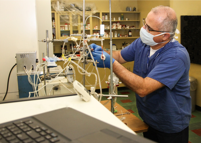
Melissa Thill is a Risk Management Project Manger at Abbott. She has prior experience in transfusion medicine and cell therapies, having worked at Fresenius Kabi. Additionally, she has expertise in bedside cell and gene therapy delivery systems gained from her time at Lupagen. She is a Marquette University BME alum with a background in systems engineering.

Learn more about Ms. Thill

Transfusion Medicine—Collection, Processing, Testing, and Administration


Taosheng Liu, PhD  March 22, 2024

Dr. Taosheng Liu is a Professor of Cognition and Cognitive Neuroscience in the Department of Psychology at Michigan State University. Dr. Liu's primary research focuses on visual selective attention and extends to related areas like visual working memory and decision making. He is interested in exploring the interconnected processes from perception to action with a focus on the computational and neural mechanisms of these psychological functions.

Learn more about Dr. Liu

Neural mechanisms of attention to visual features and objects 

It is well established that visual perception is strongly shaped by selective attention. However, how selection is achieved in the brain remains a major question in cognitive neuroscience and, in particular, the neural mechanisms underlying attention to non-spatial properties remain poorly understood. I will review recent work from our group that investigates the neural mechanisms of feature- and object-based attention, with a focus on the neural instantiation of attentional priority for these properties, how priority signals modulate behavior, and the computational principles underlying these neural representations. I will discuss how neural computations confer the brain with an efficient and robust mechanism to support flexible attentional control. 


Gopal Iyer, PhD  April 5, 2024

Dr. Gopal Iyer is an Assistant Professor in the Department of Human Oncology at the University of Wisconsin, Madison. His research seeks to decipher the dynamics of signaling bias regulated by epigenetic and phosphorylation processes. 

Learn more about Dr. Iyer

Unraveling the G-protein gamma subunit GNG2 in Lung Cancer Metastasis to the Brain 

Lung cancer metastasis to the brain remains a critical challenge in cancer treatment, with limited therapeutic options and poor patient outcomes. My lab focuses on elucidating the molecular mechanisms driving this devastating process, particularly the role of Netrins in the extracellular matrix (ECM) and their regulation by the heterotrimeric G-protein gamma subunit (GNG2). Preliminary findings have identified GNG2 as a significant regulator of lung-to-brain metastasis, suggesting its involvement in modulating the Netrin-1 and Netrin-4 pathways through focal adhesion kinase (FAK) activation. Our approach combines molecular biology, advanced imaging, and biomechanical techniques to provide a comprehensive understanding of the GNG2-FAK-Netrin axis in lung cancer metastasis. Using both in vitro and in vivo models, we investigate how alterations in GNG2 expression impact cancer cell migration and invasion within Netrin-modified microenvironments. By leveraging human cerebral organoids, we simulate the brain microenvironment to analyze tumor cell behavior under GNG2-FAK signaling and assess the biomechanical aspects of organoid invasion and ECM stiffness modulation. By integrating insights from these diverse approaches, we aim to uncover novel therapeutic targets and develop improved treatment strategies for lung cancer patients, especially those at risk of brain metastasis. This research holds the potential to significantly advance our understanding of the complex mechanisms underlying lung cancer metastasis to the brain, paving the way for more effective and personalized therapies. Our ultimate goal is to translate these findings into clinical practice, improving the lives of lung cancer patients and their families.


Shank Rao, PhD  April 19, 2024

Shashanka Rao is currently a Post-doctoral Fellow in Dr. Brandon Tefft's Cardiovascular Regenerative Engineering (CaRE) Laboratory in the Marquette-MCW Joint Department of Biomedical Engineering. He obtained his PhD from LSU Health Science Center in Shreveport, Louisiana. He specializes in redox biology in the context of cardiovascular diseases such as hypertension and atherosclerosis.  

Learn more about the CaRE Lab

Using ex-vivo siRNA approach to endothelialize commercially available vascular grafts

Myocardial infraction is a leading cause of death, and surgical intervention is often required to treat severe coronary artery disease. Synthetic vascular grafts less than 6 mm diameter fail due to unacceptable patency rates (approximately 60% at 1 year of implantation). The loss of patency is accredited to platelet activation, thrombosis, and neointimal hyperplasia. A promising strategy to improve patency is to endothelialize synthetic vascular grafts ex vivo, but previous attempts have met with modest success due to detachment of cells upon exposure to fluid shear stress. Using RNA seq approach for adherent endothelial cells (ECs), we found that fibronectin leucine-rich transmembrane protein 2 (FLRT2) was significantly downregulated in adherent ECs subpopulation. We used silencing RNA (siRNA) approach to seed FLRT2 silenced ECs onto a commercially available Goretex graft material ex vivo and subjected them to fluid shear stress of 30 dyn/cm2 for 20 minutes. We observed an increase in retention of FLRT2 silenced ECs on Goretex graft material in comparison to non-specifically targeted scramble siRNA containing ECs under shear stress.  In conclusion, we here for the first time show that siRNA approach may be a great strategy to reendothelialize commercially available vascular grafts ex vivo and that loss of FLRT2 plays an important role in EC adhesion. We would like to further investigate the molecular mechanisms underlying the role of FLRT2 in EC adhesion and use this strategy to reendothelialize commercially available synthetic grafts ex vivo and test it in our porcine models for patency.


Seminar Archive

For your convenience, the Joint Department of Biomedical Engineering provides a complete seminar archive, dating back to 2016, when the Joint Department was formed between Marquette University and the Medical College of Wisconsin.  


View Seminar Archive