The Motion Analysis Center at Shriners—Chicago features state-of-the-art facilities designed to support Motion Analysis Research including dynamic pedobarography, upper extremity analysis, segmental foot and ankle analysis, computerized balance assessment, and biplane fluoroscopy. Learn more about the facilities' featured capabilities below.
Featured Capabilities
Segmental Foot and Ankle Analysis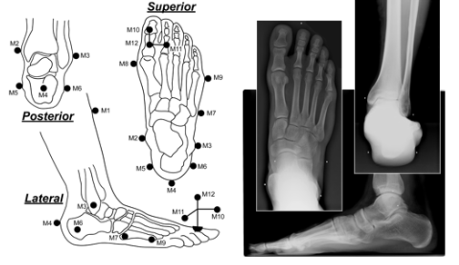
Validated for use in pediatric and adult populations, the Milwaukee Foot Model (MFM) is a segmental foot model that provides kinematic data of four foot and ankle segments: tibia, hindfoot, forefoot and hallux. Unique to the MFM is the use of weightbearing, roentgenographic offset measurements in anterior/posterior, lateral and a unique hindfoot coronal views for the purpose of neutral referencing. To improve the accuracy of captured data, the MFM uses these offset measurements to reference the positions of anatomical markers on the skin during a static calibration trial to the underlying bony anatomy. The importance of utilizing this type of technique becomes more apparent when dealing with significant foot deformities found in the pediatric populations where neutral alignment may be impossible to obtain: talipes equinovarus (clubfoot), Charcot-Marie-Tooth and planovalgus/equinovarus associated with CP.
Upper Extremity Analysis
The Motion Analysis Center analyzes movement patterns of the trunk and arms using sophisticated camera technology. This camera technology analyzes three-dimensional upper extremity joint motion (kinematics). Force transducers embedded into wheelchair push rims are used to analyze the forces acting on the arms (kinetics). The result of these non-invasive techniques is quantitative data that can be used to identify atypical upper extremity movement patterns, assist with the prescription of treatment(s), and measure the effect of treatments on upper extremity function.
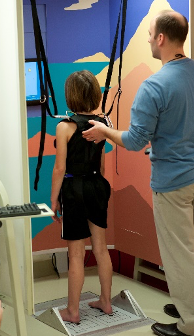 Computerized Balance Assessment
Computerized balance assessment uses sophisticated technologies to assess the sensory and motor contributions to balance deficits. The NeuroCom SMART EquiTest® system can be used to systematically evaluate how sensory information is used and how motor strategies are generated within the context of standing balance. The data generated can be used to provide a custom balance training program that targets both sensory and motor deficits.
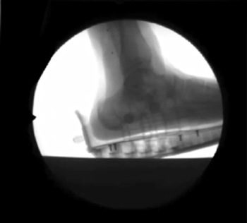
Dynamic Pedobarography
Dynamic pedobarography is a technique that measures how pressure is distributed across the bottom of the foot during walking. In the Motion Analysis Center at Shriners—Chicago, force data is recorded using a special force plate that is imbedded into the floor. This force plate generates measurements relevant to the biomechanical function of the foot. These measurements are then used to identify atypical loading patterns across the bottom of the foot.
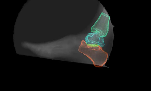
 Biplane Fluoroscopy
The Milwaukee Foot Model has proven to be useful in analyzing segmental motion of the foot and ankle; however, because the talus lacks palpable landmarks to place reliable external markers, segmental foot models—including the Milwaukee Foot Model—are unable to analyze talocrural or subtalar motion. Investigators at the Motion Analysis Center at Shriners—Chicago developed a biplane fluoroscopy system for 3D-model-based tracking of in vivo hindfoot motion during over-ground gait. The biplane system was constructed to be centered about a force plate embedded in a custom seven-meter-long walkway. Two x-ray sources and two image intensifiers have been custom mounted to the walkway with a sixty-degree angle between the sources. A CT scan of the subject is also obtained to generate volumetric models of the tibia, talus and calcaneus. The Dynamic Stereo X-Ray (DSX) Suite (C-Motion) is used to calculate joint kinematics.