Deep UV Scanning Microscopy for Breast Tumor Margin Imaging
An estimated 50-75% of women in the US who are diagnosed with breast cancer will undergo breast-conserving surgery (BCS) or lumpectomy. Women with positive margins after BCS are at 2-times the risk for cancer recurrence and are recommended to undergo additional surgery to achieve negative margins. Additional surgery is associated with significant emotional, cosmetic and financial burden for patients and their caregivers. Although several intraoperative margin assessment techniques (e.g., radiographic examination, frozen section, touch preparation and MarginProbe) are available, their accuracy is variable, and many are not routinely used, particularly in community hospitals, where most women with breast cancer receive care.
The current re-excision rate for BCS in the U.S. is 14-18% and highly variable among surgeons, ranging from zero to 92%. While several emerging imaging technologies have been proposed for intraoperative margin assessment, they are either a point- or high-resolution device with very small field-of-view that requires excessive time to scan a specimen or a wide-field device with low spatial resolution and poor sensitivity.
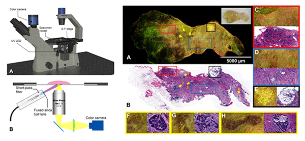
Because the size of BCS specimens varies significantly and positive margins include one or multiple sites/foci, an intraoperative device with both large margin coverage and microscopic resolution that can accurately and efficiently evaluate an entire surgical specimen is highly desirable. We have developed a deep-ultraviolet fluorescence scanning microscope (DUV-FSM) (shown in Fig. 1) that can be used for rapid and high-resolution imaging of fresh lumpectomy specimens stained with multiple fluorescence dyes. Our preliminary results show excellent visual contrast in color, tissue texture, cell density and shape between invasive lobular carcinoma (ILC), invasive ductal carcinoma (IDC), ductal carcinoma in situ (DCIS) and their normal counterparts. Statistical analysis also identified significant differences (p<0.0001) in nuclear-cytoplasm ratios (N/Cs) between invasive cancer cells and normal breast tissue elements.

Fig. 1: Intraoperative imaging of breast tumor margins during BCS including (left) Schematic of the DUV-FSM and (right) Fluorescence and H&E images of breast tissue with ILC and DCIS with adjacent adipose at the two ends; (A) fluorescence image with a specimen photo represented in the white box; (B) FFPE H&E image; (C) benign adenosis highlighted by the black box; DCIS sites indicated by yellow arrows 1, 2, 3 and 4. The zoomed-in regions at arrows 1, 2 and 3 are represented in (F), (G) and (H), respectively. The DCIS indicated by arrow 4 is not visible in the fluorescence image, likely because it is slightly below the surface.
Selected Publications
Tongtong Lu, Julie M. Jorns, Mollie Patton, Renee Fisher, Amanda Emmrich, Todd Doehring, Taly Gilat Schmidt, Dong Hye Ye, Tina Yen, and Bing Yu "Rapid assessment of breast tumor margins using deep ultraviolet fluorescence scanning microscopy", Journal of Biomedical Optics 25(12), 126501 (25 November 2020).
Bing Yu and Nirmala Ramanujam, "Quantitative Diffuse Reflectance Imaging of Tumor Margins", Chapter 17 in Biomedical Photonics Handbook, Editor-in-chief Tuan Vo-Dinh, 2nd Edition, CRC Press, 2015.
View More Biophotonics Research
 Research | Facilities | People


