New Models and Approaches to Human Analysis Modeling
Human motion analysis is focused on the study of joint motion, internal joint forces and torques, and muscle activity (electromyography or EMG). In addition to studying the major joints of the lower extremities (i.e., hips, knees and ankles), OREC's Motion Analysis Laboratories have worked to develop an accurate model of the segments of the foot. The segmental foot and ankle model is called the “Milwaukee Foot Model” (MFM) and is radiographically indexed (using X-rays) to the underlying bony anatomy. This model allows accurate motion tracking of the foot segments during ambulation in individuals (adults and children) with foot and ankle deformities. It is particularly useful for tracking segmental kinematics in children with equinovarus and planovalgus foot deformities. This includes identification of unique kinematic subgroups of children with equinovarus secondary to hemiplegic cerebral palsy. The work has also evolved to include dynamic fluoroscopy for in vivo sagittal assessment of bony hindfoot motion during gait.
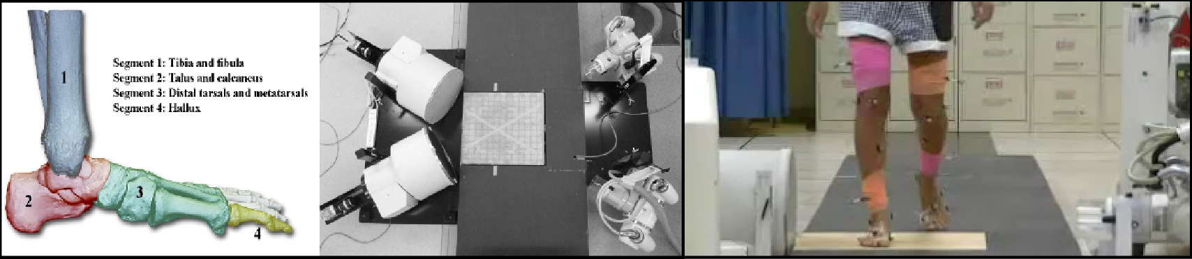 Left to Right: Milwaukee Foot Model, Biplanar Fluoroscopy System, Fluoroscopic Assessment during Gait.
Selected Publications
Ness, M.E., Long, J., Marks, R. and Harris, G.: Foot and Ankle Kinematics in Patients with Posterior Tibial Tendon Dysfunction. Gait and Posture. 27(2):331–339.
Krzak, , Corocs, D., Damiano, D., Graf, A., Hedeker, D., Smith, P., and Harris, G.: Kinematic Foot Types in Youth with Equinovarus Secondary to Hemiplegia. Gait and Posture. 41(2):402-408, 2015.
McHenry, B.D., Exten, , Long, J.T., Harris, G.F.: Sagittal Fluoroscopy for the Assessment of Hindfoot Kinematics. Journal of Biomechanical Engineering. 2016: 138 (3):034502-034502-6. doi:10.1115/1.4032445.
Amene J, Krzak JJ, Kruger KM, Killen L, Graf A, Altiok H, Smith P, Harris GF. Kinematic foot types of planovalgus feet in children with cerebral palsy, Gait & Posture. 2018 Dec 21;68:430-436.
McHenry, B.D., Kruger, K.M., Exten, E.L., Tarima, S. and Harris, G.F.: Sagittal subtalar and talocrural joint assessment between barefoot and shod walking: A fluoroscopic study, Gait and Posture, 72, 57-61, 2019.
Kruger, K., Graf, A., Flanagan, A., McHenry, B., Altiok, H., Smith, P., Harris, G. and Krzak, J.: Segmental Foot and Ankle Kinematic Differences between Rectus, Planus, and Cavus Foot Types, J Biomech, 94:180-186, 2019.
Kruger, K., Krzak, J., McHenry, B., Flanagan, A., Graf, A., Altiok, H., Smith, P., Harris, G.: Comparative Analysis of Anatomic Coordinate Systems to Calculate Hindfoot Kinematics using Biplane Fluoroscopy, Biomed Sci Instrum, 55:2, 373-378, 2019.
View more MAL Research
Get Involved!
Opportunities are available for individuals looking to get involved with OREC or the Orthopaedic and Rehabilitation Engineering communities.
View Opportunities