
Contact Us!
For more information on the Respiratory Biomechanics Laboratory, contact Dr. Guilherme Garcia.
Report a Problem
To report a problem with this website, contact BME Communications or report an accessibility issue.
In Silico Simulations | In Vitro Experiments | In Vivo Measurements
The Airway Biomechanics Laboratory uses a wide range of computational techniques, in vitro experiments, and in vivo measurements to investigate respiratory biomechanics. This diverse investigative toolset allows the Airway Lab to comprehensively investigate respiratory physiology and pathophysiology.
Computational Fluid Dynamics
Computational fluid dynamics (CFD) is a technique designed to simulate airflow, heat transfer, and the transport of airborne particles. In the Airway Lab, patient-specific models of the respiratory tract anatomy are built from computed tomography (CT) scans and magnetic resonance images (MRI).

Fluid-Structure Interaction Techniques
Fluid-Structure Interaction (FSI) techniques simulate both airflow and the movement of airway walls in response to forces generated by the airflow. This contrasts to CFD simulations, where walls are assumed to be rigid and stationary.

Computational Biology
Computational biology is a technique to simulate transport processes and chemical reactions at the cellular level.

3D-Printed Replicas

The Airway Lab uses 3D-printed replicas to quantify the pressure-flow relationships of the human upper airway.
Starling Resistor

The Starling Resistor is a model of airway collapse in obstructive sleep apnea. A collapsible silicone tube (representing the pharynx) is located between two rigid tubes (representing the nasal cavity and trachea).
Anatomically Accurate Model of Airway Collapse in Obstructive Sleep Apnea

The Airway Lab uses a combination of 3D printing and silicone molding to develop anatomically accurate models of the human upper airway with a collapsible pharynx to study the mechanics of airway collapse in obstructive sleep apnea.
Drug Induced Sedation Endoscopy (DISE)

In collaboration with Dr. Tucker Woodson (MCW Department of Otolaryngology), the Airway Lab has performed in vivo measurements of airway collapsibility in patients with obstructive sleep apnea during drug-induced sedated endoscopy (DISE).
Measuring Nasal Mucosal Temperature
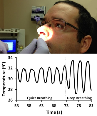
The Airway Lab has conducted In Vivo measurements of nasal mucosal temperature in healthy volunteers. Cooling of the nasal mucosa during inspiration is the one of the main mechanisms of perception of nasal airflow.
Rhinomanometry

Rhinomanometry is a technique to quantify nasal resistance to airflow. In the Airway Lab, these in vivo measurements are used to validate the pressure-flow relationship of the human nasal cavity predicted by computational fluid dynamics (CFD) models.
The team at the Airway Biomechanics Laboratory is always looking for hard-working researchers with an interest in respiratory physiology and computational modeling. For more information on becoming a member of our research team, contact Dr. Garcia.
