
Contact Us!
For more information on the Respiratory Biomechanics Laboratory, contact Dr. Guilherme Garcia.
Report a Problem
To report a problem with this website, contact BME Communications or report an accessibility issue.
Virtual Endoscopy Depth Map Videos | Select Publications
The Airway Lab is developing methods to quantify the airway cross-sectional area from endoscopy videos. As part of this project, researchers in the Airway Lab collaborated with the Marquette Visualization Lab in a pilot study which demonstrated that the airway cross-sectional area can be accurately estimated from depth maps. Virtual endoscopy videos were created from 3D models of the nasal cavity and depth maps were created in the software Blender. Clinicians in the Department of Otolaryngology at the Medical College of Wisconsin used a software application to estimate the airway cross-sectional area from the virtual endoscopy videos and depth maps. The estimated airway cross-sectional areas were in excellent agreement with measurements in the 3D models, thus demonstrating the potential of performing accurate estimates of airway cross-sectional area from depth maps. Depth maps are currently used in self-driving vehicles based on images of roads. Future studies using machine learning are needed to develop methods to compute depth maps from clinical endoscopy videos.
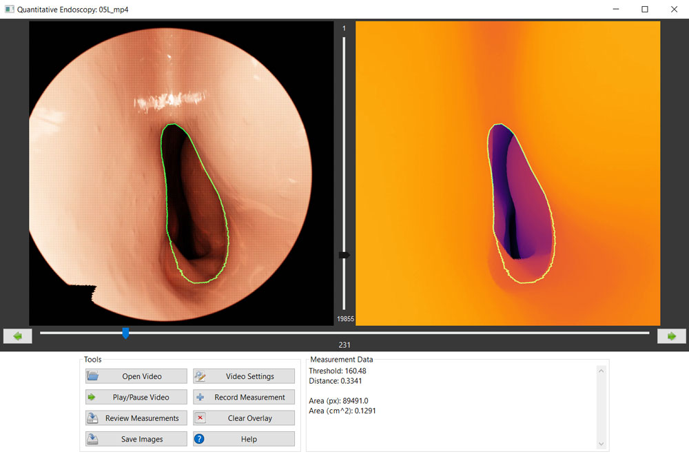
The videos below illustrate virtual endoscopies and depth maps of the nasal cavity and pharynx. The endoscope camera enters the left nostril and travels to the nasopharynx.
The following videos show a virtual endoscopy and corresponding depth map of a nasal cavity in a patient with nasal airway obstruction due to a blockage in the anterior nasal cavity.
Garcia G.J.M., Catalano D., Shum A., Larkee C.E., Rhee J.S. (2024) Estimation of nasal airway cross-sectional area from endoscopy using depth maps: A proof-of-concept study. Otolaryngology – Head and Neck Surgery 170, 1581–1589.
