
Biomedical Imaging
The Biomedical Imaging research groups in the Marquette Medical College of Wisconsin Joint Department of Biomedical Engineering focus on developing new techniques to noninvasively visualize the structure and function of living systems for clinical analysis and medical intervention. Below, learn more about the many BME laboratories contributing to the development of novel imaging technologies and other advancements in this field.
Biomedical Imaging Laboratories
 Advanced Ocular Imaging Program
The Advanced Ocular Imaging Program collaborates with researchers around the world to advance the application of non-invasive imaging techniques that examine both the structure and function of the living eye.
Learn more about the AOIP | Learn more about Dr. Carroll
 Biophotonics Laboratory
The Biophotonics Laboratory develops optical imaging techniques for minimally invasive detection and treatment of cancers and other conditions. Current projects include intraoperative assessment of breast tumor margins, cervical cancer screening, and laser ablation of liver tumors with real-time monitoring.
Learn more about the Biophotonics Lab | Learn more about Dr. Yu
 Computational Lung Physiology Laboratory
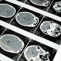
The Computational Lung Physiology Laboratory (CLPL) uses Micro-CT technology and nuclear medicine to develop experimental and computational strategies to investigate lung physiology at all levels—from molecular, to cellular, to whole-organ and body.
Learn more about the CLPL | Learn more about Dr. Audi
 Functional MRI Research Laboratory
Researchers in the Functional MRI Research Laboratory specialize in the use of MRI to assess brain function, creating imaging strategies, sequences, and instrumentation related to fMRI. This specialization may include neuroscience and vision research.
 Integrative Neural Systems Laboratory
The Integrative Neural Systems Laboratory develops techniques to fuse data sets from functional magnetic resonance imaging, electroencephalography, and functional near infrared spectroscopy to investigate the brain networks that mediate perception and action.
Learn more about the INSL | Learn more about Dr. Beardsley
 Marquette Visualization Laboratory
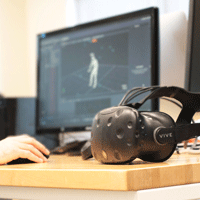
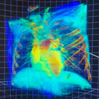
Collaborating with researchers from many disciplines, the Marquette Visualization Lab visualizes scientific data, clinical environments, and meta-data in ways that can stimulate new ideas.
Learn more about VisLab | Learn more about Dr. Cooper | Learn more about Dr. Greenberg
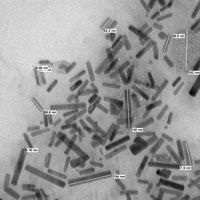 Nanomedicine & Image-Guided Interventions Laboratory
The interests of the Nanomedicine and Image-Guided Interventions Laboratory (NIGIL) include molecular imaging, photo-thermal ablation, near-infrared and shortwave infrared optical imaging and tomography systems, and contrast agent development.
Learn more about NIGIL | Learn more about Dr. Joshi
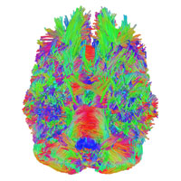
 Neuroimaging Research Laboratories
The Neuroimaging Research Laboratories focus on neural systems and neurorehabilitation imaging, including areas in MRI such as diffusion tensor imaging and tractography, and multi-modal fusion of fMRI, magnetoencephalography and electroencephalography.
Learn more about INERL | Learn more the NMCL | Learn more about Dr. Beardsley
 Ocular & Computer Vision Laboratory
The Ocular & Computer Vision Laboratory uses non-invasive imaging techniques to assess the structure and function of the living retina at both a macro and sub-micron scales.
Learn more about the OCVL | Learn more about Dr. Cooper
 Sensory Neuroscience, Attention, & Perception Laboratory
The SNAP Lab uses neuroimaging and neural stimulation methods to study the neurobiology of attention and perception in the visual, auditory, and olfactory domains while developing computational tools used to analyze data acquired through functional Magnetic Resonance Imaging, Diffusion Imaging, and Transcranial Magnetic Stimulation.
Learn more about the SNAP Lab | Learn more about Dr. Greenberg
Looking for something else?
Filter Research Themes by Research Track, or view our list of active Laboratories.
